Ongoing pulse oximetry and patient monitoring by respiratory therapists and critical care staff is key to survival and outcomes in the neonatal/pediatric intensive care unit.
By Bill Pruitt, MBA, RRT, CPFT, FAARC
Respiratory distress affects an estimated 1% of all infants in the first year of life, with significantly greater rates occurring in preemies. Pediatric patients also face risks of acute respiratory distress/failure, with prevalence being 4.6% among under-five children hospitalized for pneumonia or severe pneumonia.
Following admission, the ongoing monitoring of these patients in the neonatal/pediatric intensive care unit is key to survival and outcomes. This article will discuss effective protocols for pulse oximetry and other forms of patient monitoring in the NICU/PICU and the role of the RT and the critical care specialist.

Pulse Oximetry
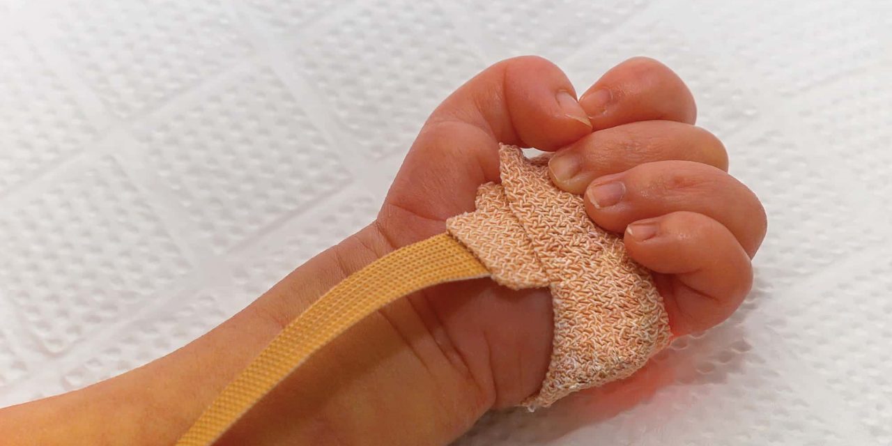
Pulse oximetry uses two different wavelengths of light (660 nm and 940 nm) that pass through the tissues from light emitting diodes to a sensor. Oxygenated and deoxygenated hemoglobin absorbs the light in different patterns and based on algorithms in the pulse oximetry microprocessor, the patterns are analyzed and real-time oxygen saturation (SpO2) can be measured.1 The process of measuring light to determine SpO2 is referred to as spectrophotometry. This noninvasive monitor has become a valuable item in detecting hypoxemia, allowing for titrating oxygen therapy and setting up/maintaining ventilatory support, etc. The light generating diodes and a corresponding light sensor are usually placed on a finger, earlobe, or toe, and the sensor can be a clip-on reusable item, or a flexible “band-aid” type designed for single-patient use. (Other locations for the sensor include the palm, foot, arm, forehead, and nasal septum).
Pulsations in blood flow are also measured in the capillary bed using the pulse oximeter using the principle of photoplethysmography. In this principle, the absorption of light increases with the increase in blood volume associated with a systolic pulse. This periodic change in blood volume/light absorption is translated into the heart rate reading.1-2 Most pulse oximeters also provide information on pulse wave activity and display the plethysmographic waves beat-by-beat.1
SpO2 readings are generally within 2% to 4% of the true saturation (arterial oxyhemoglobin or SaO2 measured by a co-oximeter from a blood sample) and calibrated to operate between a SaO2 of 70% to 100% (so SpO2 readings are not reliable below 70%).1 In the clinical setting, many clinicians consider 80% SpO2 to be the lower limit for acceptable accuracy (which corresponds to a PaO2 of about 50 mmHg at a pH of 7.40).3 Normal SpO2 is generally between 90% to 100%. Reliability is also affected by motion artifact, black or blue nail polish or artificial fingernails, poor perfusion, cold temperatures, and in patients who have dark skin tones – where the saturation may be overestimated by approximately 2%.1 Also, issues with dyshemoglobinemia (ie, the presence of carboxyhemoglobin in carbon monoxide poisoning or methemoglobin) can lead to false readings.1
The COVID-19 pandemic brought increased scrutiny of pulse oximetry and problems with obtaining trustworthy readings in black people. A paper published in 2022 in Respiratory Care describes the analysis of data from 18 studies on hypoxemia. These studies included nine Pulse oximetry brands and three types of sensors (finger clip, finger wrap, and forehead sensor).
Mixtures of air and nitrogen were administered to allow for various levels of arterial hypoxemia (measured SaO2) between 67%-100%. 3,778 data pairs were obtained from 491 subjects (849 pairs in darkly pigmented subjects, 2,929 in lightly pigmented subjects). The authors concluded that there still remains a small positive bias in Pulse oximetry readings in people with darkly pigmented skin (with the result being that some hypoxemic episodes would go undetected and untreated) and that the performance of pulse oximeters in the clinical setting is different than what is seen in the laboratory setting.4
Premature infants commonly have episodes of desaturation on Pulse oximetry monitors, which may be due to motion artifact (false hypoxemia) or a true desaturation. When a true or suspected hypoxemia occurs, the healthcare team may respond by temporarily increasing the FiO2, changing a ventilator setting, or stimulate the infant to breath by tactile stimulation if the hypoxemia is related to apnea.5 Motion artifact and suspected false hypoxemias tend to influence the team to delay or not respond, which can be detrimental to the infant. On the other hand, responding to a false hypoxemic event may expose the infant to higher FiO2 or unnecessary ventilator changes.5
In a study published in Pediatric Research in 2023, researchers examined the reliability of SpO2 during hypoxemic events associated with motion in premature infants (<28 weeks gestational age). In studying 20 infants a total of 283 hypoxemic episodes were analyzed from simultaneous readings of two pulse oximeters. Hypoxemic episodes were counted when the SpO2 < 90% for ≥ 10s. The episodes were place into four categories:
- true hypoxemia with both Pulse oximetry showing a decrease in SpO2 while only one Pulse oximetry showed evidence of motion artifact,
- false hypoxemia with one Pulse oximetry showing a decrease in SpO2 and motion artifact while the other showed no decrease in SpO2 and no artifact,
- suspected hypoxemia with both Pulse oximetry monitors showing a decrease in SpO2and motion artifact, and
- true hypoxemia – motion free (both Pulse oximetry monitors) showing a decrease in SpO2 but no motion artifact.
From their findings, the authors concluded that, “… analysis of SpO2 data collected from two pulse oximeters in the same extreme premature infant show that in the presence of motion and intermittent hypoxemic episodes, SpO2 is likely to reflect a true hypoxemia event.”5 In other words, despite motion artifact being present, the team can trust the readings from pulse oximeters and take appropriate actions.
Alarm fatigue is a recognized problem in the hospital environment. The Joint Commission (TJC) has established alarm fatigue as one of the top patient safety goals with the push to ensure that alarms are audible and response time to actionable alarms is timely.6 TJC recognizes that ICUs have “too many devices with alarms, default settings that are not at an actionable level, and alarm limits that are too narrow.”6 There are several strategies now being evaluated (and many in use) to reduce safely alarm fatigue. One of these includes using lower limits on alarms.
A study done at Children’s Mercy Kansas City (published in 2023) found that by making a few changes they were able to significantly reduce the number of nonactionable alarms (being alarms where the SpO2 was >88%). Historically the hospital had set the alarm at 90% with the goal of keeping the SpO2 >90% and had continuous monitoring as the default in ordering SpO2 in the electronic health record (EHR).
The team took action to reduce the alarm limit to 88% while maintaining the patient care goal of keeping the SpO2 >90%. Another step involved reducing the use of continuous monitoring and encouraging more use of intermittent checks on SpO2 when appropriate. This was accomplished by making the intermittent check the default order in the EHR. This study was done outside the ICU but these strategies have also been evaluated and similar approached have been successful in reducing alarm fatigue.6-7
Other Forms of Monitoring in NICU/PICU
Work is continuing in using integrated data from several monitoring systems to aid in uncovering health issues. For example, researchers combined the data gathered through pulse oximetry, near-infrared spectroscopy (NIRS), and skin temperature (ST) to develop an algorithm for early detection of sepsis in newborns.8 NIRS is an optical technique that provides information on organ oxygenation and perfusion (in particular, cerebral perfusion) by measuring regional tissue oxygen saturation and changes in blood volume. Sepsis in newborns is challenging to get an early diagnosis due to having non-specific clinical signs and symptoms. Early detection is important to help avoid the potentially serious effects of sepsis. By combining the data from these three monitoring systems, the researchers were able to increase overall accuracy of detecting sepsis by 40% and some 6 to 48 hours before the clinical diagnosis was established.8
There is growing interest in noninvasive monitoring by “wearable” means to avoid the use of wires and adhesives.9 Wired systems interfere with daily care and the benefits of skin-to-skin contact between the infant and parent (“kangaroo care,” which improves short-term physiological stability and provides long-term health benefits). Use of adhesives can cause damage to the infant’s skin with removal of various sensors, transducers, and electrodes.9 Wearable monitoring incorporates soft, flexible sensors and diagnostics into belts, caps, or harnesses to provide information such as continuous heart rate, ECG, body temperature, SpO2, and intermittent blood pressure. When the wearable monitors are combined with battery power and wireless transmission of data, wires and adhesives can be eliminated (along with less motion artifact) while providing reliable real-time information on vital signs.9
The Role of the RT and Critical Care Specialist
Respiratory therapists working in the NICU and PICU need to have a unique skill set to deal with these special patients. Infants and children are smaller and undergoing changes in their immune system as well as many other organ systems as they grow and develop.10 The various diagnosis and management of care in the NICU and PICU are often very different from what is seen in the adult ICU. The therapeutic options, equipment, and procedure are different as well, with differences found in size, complexity, and purpose compared to the adult environment. Most of the monitoring systems mentioned in this article are the responsibility of the NICU or PICU therapist for setting up, monitoring, establishing and managing the alarms, taking action when alarms are triggered, discontinuing, and cleaning/calibrating.
The healthcare team in the NICU and PICU work closely together and must communicate and cooperate well with each other, and the RTs in these areas are an integral, highly-skilled and specialized part of this team. RTs are also called on to provide education and support to family members who visit these units, to explain what is being done to care for the infant or child, and to help prepare the family for possible care needs when the patient is discharged. This specialized role is supported and credentialed by the National Board for Respiratory Care (NBRC) through the Neonatal/Pediatric Respiratory Care Specialty (RRT-NPS) credential. This credential validates the skills, knowledge, and expertise of the RT working in the NICU/PICU environment.10
The exam used to earn this credential contains 140 multiple choice questions (including 120 scored questions and 20 pre-test questions), is given over a three-hour time frame, and is open to registered respiratory therapists. As described on the NBRC website concerning the NPS exam, “Clinical competencies might range from resuscitation of the preterm neonate weighing less than one pound with underdeveloped lungs or managing a three-day-old following open-heart surgery, or a toddler with a congenital neuromuscular disorder, to an adolescent following a traumatic chest wall injury. Being able to function as a neonatal pediatric specialist is not only an intellectual accomplishment, but it also has an element of emotional intelligence as well.
The RRT-NPS needs to be able to focus on the current task to save a life and separate the sometimes difficult emotions in the moment to provide the best possible care.”10
Conclusion
Pulse oximetry has proven itself to be valuable and advancements are being made where oximetry, coupled with other monitoring systems, is increasing the value in the diagnosis and management of the complexities found in NICU/PICU cases. However, improvements in accurate measurement (particularly in darkly pigmented subjects) is needed, and the burden of alarm fatigue needs to be addressed. Wearable, wire-free monitoring and “smart” ICU monitoring systems are becoming more widely used and have improved the safety and care offered to infants and children.
Respiratory therapists working in the NICU/PICU must possess specialized skills, knowledge, and understanding to be most effective and safe as they do their jobs. Obtaining the RRT-NPS credential helps ensure that the therapists in these units have the qualities needed to be successful in these units.
RT
Bill Pruitt, MBA, RRT, CPFT, FAARC, is a writer, lecturer, and consultant. Bill has over 40 years of experience in respiratory care in a wide variety of settings and has over 20 years teaching at the University of South Alabama in Cardiorespiratory Care. Now retired from teaching, Bill continues to provide guest lectures, participates in podcasts, and writes professionally. For more info, contact [email protected].
References
- Torp K, et al. Pulse Oximetry. StatPearls. Published July 30, 2023. Accessed July 29, 2024. https://www.ncbi.nlm.nih.gov/books/NBK470348/.
- Sharma M, et al. Racial and skin color mediated disparities in pulse oximetry in infants and young children. Paediatric Respiratory Reviews. 2024 Jan 5.
- Mecham C. Pulse oximetry. UpToDate. Published May 24, 2024. Accessed August 1, 2024. https://www.uptodate.com/contents/pulse-oximetry.
- Okunlola OE, et al. Pulse oximeter performance, racial inequity, and the work ahead. Respiratory Care. 2022 Feb 1;67(2):252-7.
- Dormishian A, et al. Pulse oximetry reliability for detection of hypoxemia under motion in extremely premature infants. Pediatric research. 2023 Jan;93(1):118-24.
- Berg KJ, et al. Reducing the frequency of pulse oximetry alarms at a children’s hospital. Pediatrics. 2023 May 1;151(5):e2022057465.
- Johnson KR, et al. Reducing alarm fatigue in two neonatal intensive care units through a quality improvement collaboration. Am J Perinatol. 2018;35(13):1311–1318.
- Lungu N, et al. Enhancing Early Detection of Sepsis in Neonates through Multimodal Biosignal Integration: A Study of Pulse Oximetry, Near-Infrared Spectroscopy (NIRS), and Skin Temperature Monitoring. Bioengineering. 2024 Jul 4;11(7):681.
- Qian C, et al. Noninvasive Vital Signs Monitoring in the Neonatal Intensive Care Unit. Journal ISSN. 2024;2766:2276.
- “The Little Things Matter the Most With the NPS Credential” NBRC. Accessed August 2, 2024. https://www.nbrc.org/the-little-things-matter-the-most-with-the-nps-credential/