Optical imaging combines with computational fluid dynamics to provide precise 3D reconstructions of the upper airway, improving the detection and localization of obstructive sleep apnea.
RT’s Three Key Takeaways:
- Advanced Imaging for OSA: Swept-source optical coherence tomography (OCT) provides high-resolution, 3D imaging of the upper airway, revealing obstructions with greater precision than current diagnostic methods, study finds.
- Combination of Techniques: Researchers integrated OCT with computational fluid dynamics to visualize airflow patterns and pinpoint turbulent areas, offering a more comprehensive view of airway blockages during sleep.
- Potential Impact: Researchers say the method could change how obstructive sleep apnea is diagnosed and treated, improving surgical planning and outcomes by providing a clearer understanding of airway structure and function.
Despite advances in diagnostic tools for obstructive sleep apnea (OSA), current methods for assessing the condition remain limited, often unable to provide a complete picture of the airway obstructions that occur during sleep. This has prompted the search for a more accurate, less invasive way to diagnose OSA and guide treatment decisions.
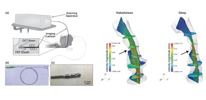
In a recent study reported in Biophotonics Discovery, researchers have explored a new imaging approach that could significantly improve how we diagnose and understand the causes of sleep apnea. The technique uses swept-source optical coherence tomography (OCT), a method typically employed in eye care, to visualize the upper airway with high precision.
By integrating a special device into the OCT system, researchers were able to extend its range and capture detailed, high-resolution images of the airway during both awake and sleep states.
Using the New OCT System
The study focused on a 28-year-old individual with sleep disorder breathing. Using the new OCT system, researchers were able to create 3D reconstructions of the upper airway, revealing significant changes between the person’s awake and sleep periods. The greatest airway obstruction was found in the oropharynx, the area at the back of the mouth, which is commonly associated with OSA.
In addition to the OCT imaging, the study incorporated computational fluid dynamics techniques to simulate airflow through the airway and pinpoint areas of turbulence, which are key indicators of obstruction. These combined techniques allowed the researchers to accurately identify where the most severe blockages occurred during sleep.
By providing clear, detailed images of the airway and airflow dynamics, this new method has the potential to revolutionize how OSA is diagnosed and treated, researchers say. By offering a more precise understanding of the airway’s structure and function, it could enhance surgical planning and improve outcomes for patients with OSA.
Photo caption: High-fidelity structural information and airflow data of a sleeping patient’s upper airway were obtained through OCT imaging and 3D reconstruction, allowing for precise identification of various anatomical sites with airflow obstruction.
Photo credit: JC Jing et al, doi 10.1117/1.BIOS.1.3.035002.