New research demonstrates that human lung tissue models can sustain immune cells for months and mount targeted responses to respiratory viruses.
RT’s Three Key Takeaways:
- Frontline Immune Response: Researchers developed lab-grown human lung models that retain resident immune cells for months and generate targeted adaptive defenses against viral infections.
- Advancing Mucosal Vaccines: Pre-stimulating the tissue models with viral fragments reduced the severity of subsequent infections, providing evidence to support the development of mucosal vaccination strategies.
- Broad Pulmonary Research Utility: The platform offers an alternative to animal testing that could help investigators study localized tissue immunity across conditions such as chronic obstructive pulmonary disease (COPD), asthma, and bacterial infections.
A new human lung model built from donated patient tissue can produce targeted immune defenses against respiratory infections, according to a study conducted by researchers at Stanford University School of Medicine and supported by the National Institutes of Health (NIH).
The findings show that lung mucosa alone can mount significant protective immune activity, suggesting that mucosal vaccination approaches targeting the respiratory tract’s frontline defenses could serve as an effective therapeutic strategy, according to the study.
“These organoids retain key immune cells and functions of lung mucosa long-term, allowing investigators to examine local tissue immunity separately from the other parts of the body’s immune system,” said John H Powers III, MD, acting director of the National Institute of Allergy and Infectious Diseases (NIAID) at the NIH, in a news release. “This platform may provide a replacement for animal models for some types of evaluations and may provide insights that tests in animal models could not capture.”
Sustaining Tissue-Resident Immune Cells
To build the platform, investigators transferred distal lung tissue samples from more than 220 patient volunteers into a specialized culture system designed to mimic the lung’s gas exchange environment. The team maintained the tissue models across a three-dimensional air-liquid interface for up to several months, during which multiple essential immune cell types survived and remained active.
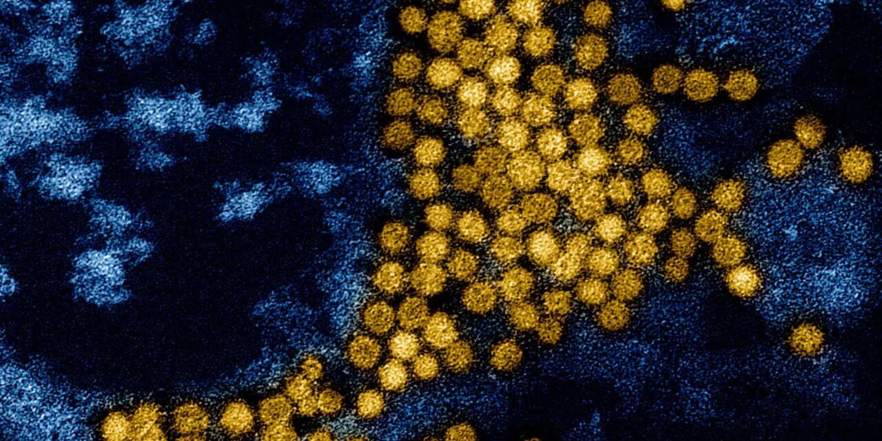
The researchers confirmed that resident T cells remained embedded within the tissue structure and retained their ability to detect respiratory viruses. When researchers infected the models with severe acute respiratory syndrome coronavirus 2 (SARS-CoV-2), the lung tissue mounted both general inflammatory reactions and virus-specific adaptive immune responses.
In an experiment simulating vaccination, the investigators exposed the tissue models to viral fragments prior to full infection. Following this pre-stimulation, the resident immune cells neutralized SARS-CoV-2 more effectively and lessened the severity of subsequent viral exposure.
“The exact role of local immunity in human lungs has long been a hotly debated subject, but we believe these organoids will be a conduit for clear answers. Already, we’ve unveiled indications that resident T cells may be much more persistent than previously thought,” said Calvin Kuo, MD, PhD, a professor of hematology at Stanford Medicine, in a news release.
Implications for Respiratory Disease and Healthcare Research
The authors reported that the results could support the design of local mucosal vaccines intended to complement or replace traditional systemic vaccines.
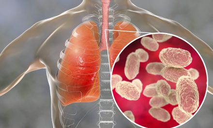
Beyond viral pathogens, investigators noted that the experimental model could help healthcare researchers examine tissue-level immune mechanisms in other respiratory conditions, including chronic obstructive pulmonary disease (COPD), asthma, and bacterial infections.