Incidence of difficult airway intubation can reach as high as 8.5% in standard practice and as high as 14% in emergency department settings.1 Achieving a successful, secure airway for these patients relies on the skills and knowledge of emergency medicine and critical care providers.
By Bill Pruitt, MBA, RRT, CPFT, FAARC
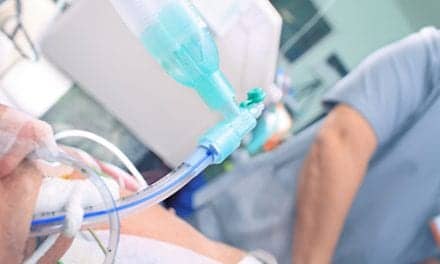
Establishing a secure, protected airway is essential care for a patient who has stopped breathing. Conditions necessitating an artificial airway may include cardiac arrest, trauma, neurological issues, drug overdose, impending cardiac or respiratory failure, etc. Intubation may be needed for an emergency, or may be an elective procedure, such as a planned surgery or invasive procedure. Temporary support can be accomplished by bag/mask ventilation, but BMV cannot be performed for prolonged periods as it can cause barotrauma from too much lung inflation and gastric insufflation which can lead to vomiting and aspiration.2
A short-term airway may also be accomplished using devices such as a laryngeal mask airway, but a secure, long-term, and protective airway is established using an endotracheal tube (or a tracheostomy tube). Having the skills and knowledge to intubate a patient is a necessary duty for emergency medicine and critical care providers. This article will explore the risk factors for difficult airway intubation in adults and discuss some of the strategies that can help achieve a successful, secure airway.

Risk Factors
There are many risk factors that can lead to difficult intubation. Most of these relate to the anatomy of the face and upper airway:3-4
- Small mouth opening
- Swollen tongue
- Short thyromental distance
- Full set of teeth with prominent incisor
- Reduced mandibular protrusion
- Reduced submandibular compliance
- Short neck
- Large neck circumference
- Limited neck extension
- Mallampati class 3 or 4
- Obesity
- Surgery or radiation-induced changes
- Trauma to the face or upper airway
- Airway secretions /blood
- Previous history of difficult intubation
- Preeclampsia
Other risk factors that may come into play include inadequate preparation (not having all equipment needed, or a needed piece of equipment is not functioning), inadequate assessment of the patient’s anatomy, not knowing the patient has a history of difficult intubation, or lack of skill in the person attempting the intubation. There is no perfect way to predict that an intubation will be difficult, but systematic evaluation of the airway using the mnemonic “LEMON” can help uncover the possibility of a difficult intubation.5
Table 1. “LEMON” method to help identify potential of difficult intubation
| L | Look for signs of trauma, neck masses, large tongue, dentures, etc. |
| E | Evaluate the 3-3-2 rule: Having less than three fingers width between the incisors, three fingers width between the hyoid bone and the mental protuberance, and/or two fingers width between the hyoid bone and the thyroid cartilage are clues that the intubation may be difficult. |
| M | Mallampati class > 3 is indicative of a difficult intubation.6 The Mallampati classification involves examination of the oropharynx with the mouth opened side and tongue extruded to assign a number from 1 to 4. 1 – The patient’s tonsils, uvula, and soft palate are completely visible. 2 – Hard and soft palate, upper tonsils, and uvula are visible. 3 – Hard and soft palate are visible, uvula is somewhat obscured. 4- Only hard palate is visible. |
| O | Obstruction or obesity that may restrict visualizing the vocal cords. |
| N | Neck mobility is limited and may contribute to difficult passage of the endotracheal tube into the trachea. |
Airway Strategies to Consider
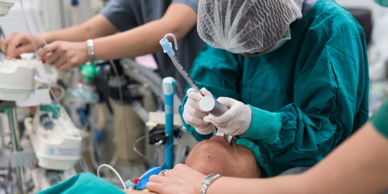
Oral Tracheal Intubation
Oral intubation is generally accomplished by two approaches:
- Direct visualization of the larynx, vocal cords, and the glottis using a Miller (straight) blade or Macintosh (curved) blade attached to a laryngoscope; or
- Video laryngoscopy, where the clinician uses a camera and observes the path on a video screen (with the same two choices for blade configuration).
Both of the above approaches enable placement for oral intubation.5 Another option involves placing the endotracheal tube over a fiberoptic bronchoscope (FOB), then visualizing and intubating the trachea using the bronchoscope to see the path to the lower airway instead of using a laryngoscope or video laryngoscope and blade. Using the FOB to intubate allows for either oral or nasal intubation.5 (See nasotracheal intubation below.)
For each intubation procedure, a primary plan and contingency plans are necessary. In the event a difficult intubation is anticipated, video laryngoscopy is often used at the onset. A 2004 publication in Anesthesia offers a flow-chart that outlines the initial plan as well as recommended backup steps in the event of unsuccessful intubation.7 It is recommended to pre-oxygenate and strive to maintain adequate oxygenation throughout the procedure.7
Primary plan (A)
Utilize effective BVM ventilation with supplemental oxygen followed by successful intubation.
If unsuccessful, resume BVM ventilation and utilize any/all of the following:
- Reposition the patient’s head and/or neck;
- Administer a paralytic medication;
- Apply cricoid pressure;
- Incorporate use of a bougie to facilitate entry into the glottic opening;
- A bougie is a long, smaller diameter tube that can pass easier through the glottic opening. The endotracheal tube is passed over the bougie once it has been placed into the glottis/trachea;
- If using direct laryngoscopy, change to a video laryngoscope.
If plan A with is unsuccessful with these added steps, move to Plan B.
Plan B
Insert a supraglottic airway (ie laryngeal mask airway) to oxygenate/ventilate and analyze the situation for options. Options include aborting the procedure (if it is elective) or use the supraglottic airway and continue with the procedure, or use the supraglottic airway as a conduit to intubate.
If these are not viable options and/or intubation is not successful, move to Plan C.
Plan C
Reattempt BVM ventilation with paralysis and ensure adequate oxygenation. Attempt to intubate using any/all of the optional steps in the primary plan. If unable to oxygenate, or intubation is unsuccessful move to Plan D. If this stage in the procedure is reached, it is often referred to as, “Can’t intubate, can’t ventilate,” or CICV.7
Plan D
Emergency front of neck access: perform a cricothyroidotomy.
For a surgical procedure, the cricothyroid membrane is located and a scalpel is used to make an incision through this membrane. Once the opening has been made by the scalpel, the index finger is inserted immediately to maintain the opening and a bougie is inserted into the opening. A 6.0 mm cuffed endotracheal tube (ETT) or a No. 4 cuffed Shiley tracheostomy tube is passed over the bougie, through the incision and into the trachea (the 6.0 mm ETT can be cut to reduce the length of tubing that extends outside of the neck). The cricothyroidotomy may also be done percutaneously.
Once an airway has been inserted, the balloon is inflated to provide a seal and allow for positive pressure ventilation through the airway. There are kits available that contain the necessary equipment to perform this procedure (eg, the Cook Melker kit) or a prefabricated percutaneous device (eg, Quicktrach II).
Standard PPE precautions should be used to protect against blood and body fluid exposure—including gloves, face mask, protective eye shield, gown, and shoe covers. This procedure should only be performed by a skilled clinician who has received formal training.8
Rapid Sequence Intubation (RSI)
In an emergency, airways are often established using rapid sequence intubation (RSI) to facilitate the procedure. This approach uses a combination of sedative and paralytic medications that have a quick onset and short duration, and are administered within a short time frame (ie, within 30 seconds). The RSI approach has been shown to increase the success of intubation on the first attempt and reduce the risk of aspiration. Since the patient is paralyzed and sedated, ventilation/oxygenation is critical and good BVM technique must be utilized to avoid hypoxia.5
Nasotracheal Intubation
Another option in cases of difficult intubation is nasotracheal intubation. The nasotracheal approach may be most useful in cases where the patient has a restricted mouth opening (ie, temporomandibular joint ankylosis or maxillofacial surgery), damaged teeth, excessive bleeding, damage in the upper airways, or where alternative supportive equipment is unavailable (ie, no access to video laryngoscope, fiberoptic bronchoscope, etc.).9 This approach is frequently used when intubating outside a hospital, such as paramedics working in the field.
The nasotracheal path is prepared by administering mucosal vasoconstrictors and local anesthetic agents. With the head and neck in the “sniffing position,” a lubricated ETT is advanced into the nose and down the nasopharynx. In a breathing patient, as the ETT approaches the epiglottis and glottis, breathing sounds can be heard through the ETT and increase in strength. During inspiration, the tube is advance through the glottis and into the trachea.9
Adding the Beck Airway Airflow Monitor (BAAM) whistle (Life Medical Supplier USA), can improve odds of nasotracheal intubation success. The device is a simple red cap that fits over the distal end of the ETT and creates a whistle with each breath that increases with intensity as the ETT approaches the glottis.10
Magill forceps can also be used. During nasotracheal intubation, the ETT may move into the esophagus rather than the glottis. Inserting the Magill forceps into the open mouth to grab the tip of the ETT as it passes through the back of the oropharynx allows the clinician to guide the tube successfully into the glottis.
Confirming Correct Airway Placement
For any intubation method, correct placement of the ETT must be confirmed. Proper tube position can be confirmed using auscultation, capnography, and radiography.11 Skin integrity is checked, and the patient’s facial and neck skin are cared for appropriately.11 Once confirmed, the ETT must be secured to prevent unplanned extubation.
Documenting the procedure in the patient’s medical record is imperative, including the challenges to intubation and the methods used to confirm ETT placement.
Conclusion
When intubating any patient, the healthcare team must be prepared for difficult airways and be armed with protocols for success. Proper equipment is essential, and the clinician (and team) must have the skills to perform all intubation contingencies, confirm airway placement, secure the tube, and document the procedure.
RT
Bill Pruitt, MBA, RRT, CPFT, FAARC, is a writer, lecturer, and consultant. He has over 40 years of experience in respiratory care and has over 20 years teaching at the University of South Alabama in the department of Cardiorespiratory Care. After retiring from teaching, he continues to provide guest lectures and write professionally. For more information, contact [email protected].
References
- Waheed S, et al. Research Waheed, S., Razzak, J.A., Khan, N. et al. Derivation of the Difficult Airway Physiological Score (DAPS) in adults undergoing endotracheal intubation in the emergency department. BMC Emerg Med 24, 40 (2024). https://doi.org/10.1186/s12873-024-00958-3
- Bucher JT, Vashisht R, Ladd M, et al. Bag-Valve-Mask Ventilation. [Updated 2023 May 21]. StatPearls Publishing; 2025 Jan. https://www.ncbi.nlm.nih.gov/books/NBK441924/
- Traylor B, McCutchan A. Unanticipated Difficult Intubation in an Adult Patient. Stat Pearls. Updated Jan 2023. https://www.ncbi.nlm.nih.gov/books/NBK572134/.
- Joffe AM, Aziz MF, Posner KL, Duggan LV, Mincer SL, Domino KB. Management of Difficult Tracheal Intubation: A Closed Claims Analysis. Anesthesiology. 2019 Oct;131(4):818-829
- Alvarado A, Panakos P. Endotracheal Tube Intubation Techniques. Stat Pearls. Updated July, 2023. https://www.ncbi.nlm.nih.gov/books/NBK560730/
- Summer J, Wright H. Mallampati Score and Predicting Sleep Apnea. Sleep Foundation. Updated October 16, 2023. https://www.sleepfoundation.org/sleep-apnea/mallampati-score
- Henderson JJ, Popat MT, Latto IP, Pearce AC. Difficult Airway Society guidelines for management of the unanticipated difficult intubation. Anaesthesia. 2004 Jul;59(7):675-94.
- Sakles J. Emergency cricothyrotomy (cricothyroidotomy). UpToDate. Updated April 2024. Accessed Jan 2025. https://www.uptodate.com/contents/emergency-cricothyrotomy-cricothyroidotomy
- Yoo H, Choi JM, Jo JY, Lee S, Jeong SM. Blind nasal intubation as an alternative to difficult intubation approaches. Journal of dental anesthesia and pain medicine. 2015 Sep 1;15(3):181-4.
- Zhang J, Lamb A, Hung O, Hung C, Hung D. Blind nasal intubation: teaching a dying art. Canadian Journal of Anesthesia/Journal canadien d’anesthésie. 2014 Nov;61:1055-6.
- Hatlestad D. Securing the Endotracheal Tube. RT: For Decision Makers in Respiratory Care. 2000. https://respiratory-therapy.com/department-management/clinical/securing-the-endotracheal-tube/