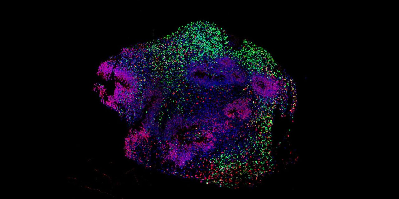
Researchers at UCLA use human stem cell-derived models to replicate the brain wave patterns produced by general anesthesia.
RT’s Three Key Takeaways:
- Human Brain Models: Researchers demonstrated for the first time that three-dimensional human stem cell-derived brain assembloids can reproduce the electrical changes seen during general anesthesia.
- Synchronized Neural Rhythms: Exposure to propofol caused individual neurons to quiet down while synchronizing network activity to create broad, slow brain waves, confirming that a minimal cortical circuit can generate these signatures without input from the thalamus.
- Broader Clinical Applications: The model offers a platform to study why patients differ in anesthetic sensitivity and to evaluate conditions that disrupt brain networks, including traumatic brain injury, coma, and epilepsy.
UCLA researchers have shown for the first time that human stem cell-derived brain assembloids can recreate the electrical shifts observed during general anesthesia, according to a study published in the British Journal of Anaesthesia.
The tiny, three-dimensional models recreate simplified human brain circuits, providing a functional platform to examine how molecular changes within individual cells generate large-scale electrical rhythms across the brain. For healthcare clinicians in critical care and surgical settings, understanding these network dynamics remains vital for managing sedation and monitoring patient consciousness.
“We’ve known what these drugs bind to for a long time, and we’ve known what happens at the level of the whole brain,” said Daniel Toker, first author of the paper and a project scientist in the laboratory of senior author Ranmal Samarasinghe. “What we’ve been missing is a human model that lets us connect those two scales.”
Recreating Cortical Circuitry
Created from induced pluripotent stem cells, the assembloids combine excitatory neurons, inhibitory neurons, and glial cells, according to the study. Together, these cellular elements form interconnected networks that generate coordinated rhythms resembling those in the living human brain.
To evaluate the platform, the research team exposed the models to the anesthetic propofol. The assembloids produced the broad, slow brain waves characteristic of general anesthesia on an electroencephalogram (EEG), even as the electrical activity of individual neurons decreased significantly.
“It’s a little counterintuitive,” said Toker. “Individual neurons become less active, but the overall brain waves become larger, because many neurons begin changing their activity in synchrony.”
The researchers confirmed that this response relied on established biological mechanisms: blocking the specific receptors targeted by propofol eliminated the slow-wave effect, and simpler cell models lacking inhibitory neurons could not generate the waves, the study reported. Additionally, because the assembloids contained no thalamus, the findings demonstrate that a minimal cortical network is sufficient to generate these slow-wave signatures on its own, resolving a longstanding scientific debate.
Implications for Clinical Research and Sedation
Beyond general anesthesia, researchers noted that the platform could help screen candidate drugs and investigate why individual patients differ in their sensitivity to anesthetics, including instances of accidental awareness during surgery.
“We’re excited about these models’ potential not just for studying anesthesia, but as a tool we can use more broadly to understand how brain networks become disrupted in other disorders that profoundly alter brain dynamics like traumatic brain injury,” said Ranmal Samarasinghe, assistant professor of neurology and member of the UCLA Broad Stem Cell Research Center.
By offering a controlled human model to investigate how brain networks transition between electrical states, researchers aim to advance the study of conditions ranging from coma and traumatic brain injury to epilepsy, the authors stated.
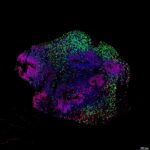
Image: Representative image of a developing human cortex-like organoid. In this study, these organoids were later fused with interneuron-producing organoids to form the assembloids used for electrophysiological recordings. Image credit: Immunohistochemistry by Emily Duncan and imaging by Dr. Adrianna Carrasco of the Samarasinghe Lab