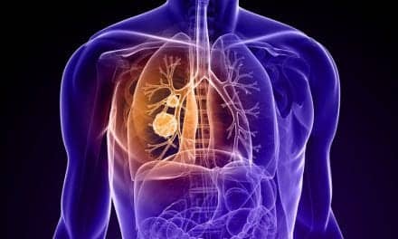

A new noninvasive approach combines imaging analysis and blood testing to evaluate indeterminate lung nodules.
RT’s Three Key Takeaways:
- Award for Early Detection: A multidisciplinary team at UCLA received a $1 million grant from the LUNGevity Foundation and the Rising Tide Foundation for Clinical Cancer Research to develop a noninvasive diagnostic method for evaluating lung nodules.
- Combined Diagnostic Strategy: The three-year project integrates computed tomography (CT) radiomics with blood-based cell-free DNA methylation testing to assess lung cancer risk more accurately.
- Clinical Validation in Patients: Researchers will validate the approach in 500 patients across UCLA Health and Veterans Affairs medical centers to reduce unnecessary invasive biopsies and surgeries.
A multidisciplinary research team from UCLA Health has received the inaugural Early Detection Award from the LUNGevity Foundation and the Rising Tide Foundation for Clinical Cancer Research, according to the university.
The $1 million award will support the development of a noninvasive diagnostic method to determine whether indeterminate pulmonary nodules detected on lung CT scans are malignant.
The initiative brings together specialists in cancer biology, pulmonary medicine, computational imaging, artificial intelligence, and molecular diagnostics. Led by Steven Dubinett, MD, the research group also includes Ramin Salehi-Rad, MD, PhD, Xianghong J. Zhou, PhD, William Hsu, PhD, and Linh M. Tran, PhD.
Addressing the Clinical Challenge of Pulmonary Nodules
More than 1.5 million individuals across the US are diagnosed each year with an indeterminate pulmonary nodule. Although the majority of these nodules turn out to be benign, diagnostic uncertainty frequently leads to invasive procedures that prove unnecessary, while other patients experience delays in diagnosis and timely therapy.
Because lung cancer remains the leading cause of cancer-related mortality in the US, early and precise detection remains critical to improving long-term clinical outcomes in healthcare systems. The UCLA team intends to equip clinicians with a reliable assessment tool to identify high-risk patients who require swift intervention while sparing low-risk patients from avoidable procedural risks.
Integrating Imaging and Molecular Biomarkers
Over the course of the three-year study, the investigators will pair advanced CT scan analysis with a blood-based test. The protocol integrates radiomics—which applies computational algorithms to extract quantitative features from medical scans—with the evaluation of cell-free DNA methylation in peripheral blood.
The investigators plan to validate this dual testing strategy in 500 patients undergoing care within UCLA Health and Veterans Affairs medical centers. The primary objective is to evaluate whether coupling radiomic and molecular biomarkers can enhance risk stratification, support clinical decision-making, and curb unnecessary tissue biopsies and surgeries.
“This award recognizes the power of team science and the value of bringing together experts from multiple disciplines to tackle a critical problem in lung cancer detection,” said Dr Dubinett, dean of the David Geffen School of Medicine at UCLA, associate vice chancellor for research at UCLA, and an investigator at the UCLA Health Jonsson Comprehensive Cancer Center, in a news release. “By integrating advances in imaging science, artificial intelligence, and molecular diagnostics, we hope to improve assessment of pulmonary nodules and help ensure that patients receive the right care at the right time.”