
PICU specialists at Children’s Memorial Hermann share four case studies of patients receiving ventilatory support who benefitted from the hospital’s adoption of transcutaneous CO2 monitoring and the continuous visibility it provides.
By iAngel Haver, RRT, RRT-NPS; iAlvaro Coronado Munoz, MD; iKonstantinos Boukas, MD; and iRick Hinojosa, BS, RRT, RRT-NPS, CPFT
This content is sponsored by Sentec AG. Join author Angel Haver on Wednesday March 13th, 2024 at 12:00pm ET for a live discussion of tcPCO2 monitoring in the Children’s Memorial Hermann PICU. Register here. (CRCE application pending.)
Summary
As care teams in pediatric intensive care units (PICUs) have increasingly turned to noninvasive and specialty modes of ventilatory support for their respiratory patients, continuous visibility to ventilation status through carbon dioxide (CO2) levels has become vital.
The PICU at Children’s Memorial Hermann is no exception, and has adopted transcutaneous CO2 monitoring (Sentec, Therwil, Switzerland) to meet the need for continuous visibility, particularly in patients supported on noninvasive ventilation (NIV) and specialty ventilation. The case studies explored in this paper demonstrate the impact of this technology in supporting earlier interventions for respiratory patients in the PICU, as well as its utility under a variety of challenging circumstances.
Facility & Patient Population Overview
Children’s Memorial Hermann Hospital, housed within the flagship medical center of the award-winning Memorial Hermann Health System in Houston, Texas, is a busy pediatric hospital that handles 16,000 emergency center visits, delivers more than 4,600 babies, and performs 6,000 surgeries each year. It is home to more than 40 specialty programs including pediatric trauma and cardiac units, as well as the neonatal intensive care unit.
Like pediatric ICUs across the world, Children’s Memorial Hermann admits a large number of children annually who require respiratory support. These patients are increasingly common; in fact, one study found that as many as one in every three patients admitted to the PICU require ventilatory support.1
CO2 Monitoring in the PICU
Over the last 20-30 years, preferred methods of respiratory support in the PICU have undergone a significant paradigm shift toward noninvasive methods of ventilation. Data indicate that use of mechanical ventilation has dropped over time, as use of noninvasive methods like nCPAP have grown dramatically.2
Although our case studies primarily feature patients who are mechanically ventilated, this context is valuable in understanding the important role of carbon dioxide (CO2) monitoring in our PICU. Our practice has similarly increased the use of noninvasive ventilation modalities for our patients, particularly those who are difficult to manage on mechanical ventilation, like those with asthma or bronchiolitis.
Regardless of ventilation modality, CO2 is an important parameter to monitor closely, as demonstrated by the included case studies below of patients with severe chronic lung disease, acute respiratory failure, and acute respiratory distress syndrome. In addition to offering an indication of ventilation status, CO2 monitoring also helps to avoid hypercapnia and hypocapnia—both of which have been associated with increased mortality risk.3-5 And in the case of hypercapnia in particular, caregivers must also be concerned with poorer outcomes, higher costs, and longer hospital stays.6
Our team utilizes several different tools to monitor CO2 levels in their patients, including arterial blood gases (ABG/PaCO2), end-tidal CO2 monitoring (etCO2), and transcutaneous CO2 monitoring (tcPCO2).
Arterial Blood Gases
ABGs often guide clinical decision-making in our PICU. However, frequency of these draws varies depending on patient type and provider determination of need on a case-by-case basis. In recent years, reliance on blood gases has decreased, largely due to increasing trust in continuous, noninvasive methods of monitoring.
Performing ABGs in the PICU has drawbacks; some inherent to the procedure and others related to workflow inefficiencies. For example, blood draws can cause anemia and therefore the need for transfusions, particularly in the most fragile, needy PICU patients.
Further, the time it takes to get results from a blood gas can mean hidden costs and potentially even delays in patient care, especially related to the need for increased respiratory support.7 Although ABGs are the gold standard in accuracy, they provide only a point-in-time measurement, which can sometimes result in misrepresentation of patient course.
etCO2 Monitoring
End-tidal CO2 has broad applications across our PICU. All ventilated patients have historically been monitored with etCO2 measurements, with exceptions for neonatal patients for whom the technology is typically unreliable. It has further usage in consciously sedated patients and for Extubation Readiness Trials (ERT) to determine adequate ventilation of patients. End-tidal CO2 has also become a standard of care here for monitoring the quality of chest compressions when staff is performing CPR.
In addition, etCO2 is a recommended tool for the monitoring and management of dead space in pediatric acute respiratory distress syndrome patients being managed on mechanical ventilation.10 Together with PaCO2, using etCO2 to calculate dead space can help clinicians assess the severity of lung disease.
However, for many patients, end-tidal monitoring is infeasible or inaccurate. (See Figure 1.) While our team will often continue monitoring with end-tidal CO2 for trending purposes, we are vigilant for circumstances that require an alternate monitoring method.
| Figure 1. When is etCO2 infeasible?8-9 |
| • Noninvasive ventilation (NIV) • High-frequency ventilation (HFV) • Patients with ventilation perfusion (V/Q) mismatch • Patients with severe bronchopulmonary dysplasia (BPD) • Patients with persistent pulmonary hypertension of the newborn (PPHN) • Patients with airway malacia |
Transcutaneous CO2 Monitoring
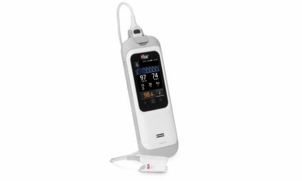
Monitoring CO2 using tcPCO2 measurements was introduced to the Children’s Memorial Hermann Hospital PICU in 2020. Previously used by Dr. Alvaro Coronado Munoz (who joined Children’s Memorial Hermann in 2017) during his time at the University of Miami, he and others on the staff championed its introduction.
As the broader PICU team’s trust has grown in the transcutaneous monitor (Sentec AG, Therwil, Switzerland), its use cases have continued to expand.
This method is used for a variety of patients in our PICU, including:
- Patients on high-frequency oscillating ventilation;
- Patients on extracorporeal membrane oxygenation (ECMO), especially those weaning on or off ;
- High-acuity patients on a ventilator who require high airway pressure and for whom V/Q mismatch causes etCO2 inaccuracies;
- Those on high-flow nasal cannula (HFNC) or NIV for whom ventilatory status and the potential for intubation is a concern;
- Asthmatic patients on BiPAP; and
- Croup patients on Heliox (in order to assess successful bypassing of upper airway obstruction).
Hypercapnia can have serious implications for PICU patients; data indicates that 26% of pediatric NIV failure cases involve hypercapnia.6 Transcutaneous CO2 monitoring can allow clinicians to perform earlier interventions for patients who are not intubated, as well as for those who are on speciality forms of ventilation or facing other conditions that make end-tidal CO2 monitoring infeasible.
The Quality Improvement Committee at Memorial Hermann set goals tied to improving patient outcomes by targeting earlier interventions. Our PICU team is working to meet these objectives by targeting CO2 for early initiation of noninvasive respiratory support to avoid intubation, as well as to optimize mechanical ventilation and execute lung-protective strategies.
| Why transition to APRV? |
| The main reason in our PICU for switching the ventilator mode to APRV is to improve oxygenation using the ventilator pressures consistent with lung protection (to avoid overdistension). Other lung-protective strategies at Children’s Memorial Hermann include: • Permissive hypercapnia (to deliver small tidal volumes and lower airway pressure) • Increased PEEP (to prevent atelectrauma and reduce potential for shear stress) • Plateau pressure assessment and ventilatory strategy adjustment to keep values below 28 cmH2O |
Case Studies
Case Study 1
Infant, approximately 18-months-old; male; severe chronic lung disease, pulmonary hypertension, and tracheobronchomalacia.
This patient was transferred to Children’s Memorial Hermann for a shunt revision, along with a history of bronchopulmonary dysplasia secondary to prematurity. Patient history also included severe tracheobronchomalacia (collapse of the trachea and main bronchus during inhalation), which required tracheostomy and long-term mechanical ventilatory support.
Following the shunt revision, this patient’s status remained poor. During their prolonged stay in our PICU, this (approx.) 2-year-old experienced multiple infections (tracheitis, line infections, and pneumonia).
In keeping with our typical protocols, this patient was initially monitored with end-tidal CO2. However, flow limitations caused by the collapse of this patient’s airway, chronic lung disease, and secretions from multiple episodes of tracheitis led to inaccurate etCO2 readings. Further, this patient frequently had CO2 levels that exceeded 90 mmHg, which the end-tidal CO2 monitor was unable to read.
The inline CO2 adapter increased dead space in the circuit, which the patient did not tolerate; and the patient’s capacity to trigger supported breaths was impacted regardless of the trigger sensitivity setting. As a result of these limitations and the need for continuous monitoring of this patient’s CO2, staff decided to place a transcutaneous CO2 monitor. This quickly resulted in more accurate correlations with PaCO2 measurements, even during spikes in CO2 levels.
Throughout bouts of respiratory acidosis, sepsis with peripheral hypoperfusion, and septic shock, the transcutaneous readings continued to correlate well even at values as high as 90 or even 120 mmHg. In these situations, the team was able to quickly perform interventions to compensate for worsening ventilation status. These spikes were typically the result of severe tracheobronchomalacia, and were exacerbated by episodes of infection.
The severity of this infant’s tracheobronchomalacia meant our team used transcutaneous CO2 monitoring on this patient during their entire prolonged stay in our PICU. Even given this long-term usage, this patient had no skin injury from the sensors.
For this challenging patient, accurate continuous CO2 monitoring was necessary, and etCO2 presented limitations. This case reflects the utility of the transcutaneous CO2 monitor in patients with severe chronic lung disease with high CO2 levels. Transcutaneous CO2 monitoring helped our team make decisions about ventilatory settings, ventilator modalities, and even the delivery of paralytic agents to gain control of the patient’s ventilatory status when necessary.
Case Study 2
Infant, approximately 14-months-old; female; worsening acute respiratory failure.
This patient was admitted to the PICU with worsening acute respiratory failure after being transferred from the emergency room for exacerbation of reactive airway disease caused by viral bronchiolitis. They were placed on CPAP, and initial transcutaneous CO2 readings were trending between 50-60 mmHg. These readings were confirmed by an arterial blood gas.
The patient continued to have increased work of breathing, and bedside staff soon noticed the tcPCO2 trending upward. Given the increasing CO2, values and worsening patient condition, the team decided to increase respiratory support. The patient was transitioned to BiPAP and the tcPCO2 reading improved.
The additional support was prompted by early detection of worsening respiratory status in this patient as detected by the tcPCO2 trend. Importantly, the proactive decision to move the patient to BiPAP was made without further invasive or painful testing.
Patient condition subsequently improved as her respiratory status stabilized on BiPAP.
Utilizing tcPCO2 monitoring can help pediatric critical care teams perform prompt interventions in patients who do not have arterial lines or means to measure etCO2. Transcutaneous monitoring in this patient’s case allowed the team to respond quickly, potentially avoiding further respiratory fatigue which could have ultimately led to respiratory failure and intubation. During this patient’s stay, the team was able to avoid intubation altogether.
Case Study 3
Teenager; approximately 15-years-old; female; severe acute respiratory distress syndrome; transferred to HFOV.
In this rare case, this previously-healthy teenage female presented with sinusitis caused by a respiratory infection. This seemingly minor infection progressed, causing the patient to become septic and subsequently develop ARDS, which required intubation.
During her PICU stay, she experienced septic shock and acute hypoxemic respiratory failure while on mechanical ventilation. As her lung compliance worsened, she developed severe acute respiratory distress syndrome with an oxygenation index of 35 and a PaO2/FiO2 ratio of 85, resulting in severe respiratory acidosis.
The patient was supported on conventional mechanical ventilation using lung-protective strategies, tolerating a pH of 7.2. As this patient’s condition worsened, ventilation perfusion mismatch increased, and etCO2 readings began to become inconsistent with PaCO2 measurements; as a result, the team decided to place a transcutaneous monitor.
Unfortunately, the ventilator settings required to appropriately oxygenate and ventilate the patient soon became exceedingly high, prompting the team to transition this patient to airway pressure release ventilation (APRV).
When APRV was initiated, the PaCO2 was measured at 70-80 mmHg with a pH of 7.22-7.3. Over the next few hours of treatment, the bedside respiratory therapist reported transcutaneous CO2 readings rising to 90-100 mmHg. An ABG was drawn, confirming the accuracy of the reading and revealing a pH level of 7.1 and worsening oxygenation.
Due to the patient’s worsening ventilation status, the team transitioned the patient to HFOV. Because etCO2 is not measurable while on HFOV, the team continued to monitor CO2 transcutaneously. The tcPCO2 values correlated accurately with PaCO2 measurements, which allowed the caregivers to feel confident making real-time changes to the oscillator when ventilation (as indicated by the tcPCO2 value) was impaired.
CO2 goals were set to adjust amplitude and frequency settings on the oscillator, and by closely monitoring the patient and managing CO2 levels, the team was able to prevent complications like respiratory acidosis and further lung injury. Transcutaneous CO2 measurements also reduced the need for frequent, painful arterial blood gas draws during the patient course.
Close monitoring of CO2 levels of this patient allowed our team to safely escalate respiratory support, as well as to make timely titrations to avoid any further, or potentially long-lasting, damage to this patient’s lung tissue. This patient made a full recovery and was discharged without any respiratory compromise or chronic respiratory issues caused by time on mechanical ventilation.
| What Is Apnea Testing? |
| Apnea testing is part of the clinical assessment of brain death to prove the absence of respiratory control system reflexes in the brainstem when it is triggered to breathe. “The outcome of an apnea testing is very straightforward. A negative test is defined by any spontaneous respiratory efforts in response to hypercapnic/acidotic stimulation; a positive test is the absence of any respiratory activity under these conditions.” 9 |
Case Study 4
Pediatric, approx. 9-10 years old; male; trauma due to motor-vehicle collision; ventilated; required apnea brain-death test.
In order to diagnose brain death, an apnea test is performed by removing ventilatory support and assessing if spontaneous breathing is triggered at certain CO2 levels. Blood gases are generally obtained serially to detect changes in CO2, often at 5 and 10 minutes.
The duration of this test can vary, depending on the patient and hospital protocols; the test can continue as long as the patient can tolerate being off the ventilator without desaturation or hemodynamic changes, even up to 15-20 minutes. The test ends either when a) the patient breathes, resulting in a negative test for brain death, or b) the PaCO2 levels rise to >60 mmHg11 and greater than 20 mmHg from baseline. If the patient does not breathe at this point, this indicates a positive test result for brain death, which must be confirmed with a blood gas.
“Current guidelines for apnea testing state that measurement of the blood gas should be conducted after approximately 8-10 min;” however, “apnea testing may be continued until the arterial blood gas results are noted, as long as the patient remains hemodynamically stable and with an adequate SpO2.” 11
Our PICU’s protocols reflect the above guidance. However, the team is also able to use tcPCO2 monitoring to help reduce both the number of blood draws and, in some cases, the length of this very stressful procedure. Drawing an ABG when the tcPCO2 value reflects that the apnea test parameters have been met can mean a reduction in intermittent gases drawn while waiting for the CO2 value to rise, and also the duration of the test if tcPCO2 indicates the parameters are met quickly. Both of these are valuable to everyone in the room: the patient, the care team, and, often, the patient’s family. In this way, tcPCO2 acts as a guide for our team and a way to minimize stress during these critical moments.
In this case, an apnea test was ordered for a young male patient who was admitted with a traumatic brain injury resulting from his involvement in a motor vehicle crash. A tcPCO2 sensor was applied as an extra monitoring tool. The team utilized the transcutaneous CO2 value as a reference to detect any increases in CO2. These values enabled the team to draw the blood gas at four minutes for this patient, decreasing the duration of the apnea test and allowing the patient to return to the ventilator before they became hemodynamically unstable. Unfortunately, in this case, apnea was confirmed in this patient. Although this is never the desired outcome, monitoring CO2 continuously allowed our team to obtain the results of this test quickly, creating a more controlled, calm environment in a highly sensitive scenario.
This technique is supported by research in Respiratory Care, which states, “Transcutaneous CO2 monitoring has been reported to prevent excessive hypercarbia/respiratory acidosis resulting from the apnea testing procedure.”11
Conclusion
The PICU at Children’s Memorial Hermann Hospital is continuing to implement transcutaneous CO2 monitoring. Together with existing technologies and practices, transcutaneous CO2 monitoring is an important aspect of care for patients like those on speciality ventilation or NIV, or for whom end-tidal CO2 monitoring cannot be relied upon. Our PICU team expects that use of transcutaneous monitoring will expand as more data is gathered about where it can be most helpful in supporting the care of respiratory patients beyond the use cases explored here.
Angel Haver, RRT, RRT-NPS is clinical coordinator for PICU; Alvaro Coronado Munoz, MD is Pediatric Critical Care physician; Konstantinos Boukas, MD is Pediatric Critical Care physician; and Rick Hinojosa, BS, RRT, RRT-NPS, CPFT is RT manager at Children’s Memorial Hermann Hospital. For more information, please contact [email protected].
Author Affiliations
iChildren’s Memorial Hermann Hospital (Houston, Tex)
References
- Farias JA, et al. What is the daily practice of mechanical ventilation in pediatric intensive care units? A multicenter study. Intensive Care Medicine. 2004.
- Essouri S, et al. Improved clinical and economic outcomes in severe bronchiolitis with preemptive nCPAP ventilatory strategy. Intensive Care Medicine. 2014.
- Madotto F, et al. Patterns and Impact of Arterial CO2 Management in Patients With Acute Respiratory Distress Syndrome: Insights From the LUNG SAFE Study. CHEST. 2020.
- Fuller BM, et al. Partial pressure of arterial carbon dioxide and survival to hospital discharge among patients requiring acute mechanical ventilation: A cohort study. J Crit Care. 2017.
- Nin N, et al. Severe hypercapnia and outcome of mechanically ventilated patients with moderate or severe acute respiratory distress syndrome. Intensive Care Med. 2017.
- KID database documentation. 2019.
- Emeriaud G, et al. Executive Summary of the Second International Guidelines for the Diagnosis and Management of Pediatric Acute Respiratory Distress Syndrome (PALICC-2). Pediatric Critical Care Medicine. 2023.
- Tobias JD, et al. Noninvasive monitoring of carbon dioxide during respiratory failure in toddlers and infants: end-tidal versus transcutaneous carbon dioxide. Anesthesia & Analgesia. 1997.
- Berkenbosch JW, et al. Transcutaneous carbon dioxide monitoring during high-frequency oscillatory ventilation in infants and children. Critical Care Medicine. 2002.
- Storre, JH, et al. Monitoring of patients receiving mechanical ventilation. Pneumologie. 2014.
- Scott J, et al. Apnea Testing During Brain Death Assessment: A Review of Clinical Practice and Published Literature. Respiratory Care. 2013.


Hello, how should we charge for the Transcutaneous CO2 Monitoring? What are the recommended CPT codes? Thank you!