The scanning method reveals how air moves in and out of the lungs during breathing in patients with asthma, COPD, and those who have received a lung transplant.
RT’s Three Key Takeaways:
- An MRI using inhaled perfluoropropane gas allows for real-time monitoring of lung function in patients with asthma, COPD, and lung transplant recipients.
- The technique can reveal areas of the lungs with poor ventilation, helping assess the effects of treatments like bronchodilators and track lung function changes in transplant patients.
- Researchers say the scan method’s sensitivity could lead to earlier detection of lung function decline, improving care and supporting the development of new treatments for respiratory conditions.
A new method of scanning lungs is able to show the effects of treatment on lung function in real time and enable experts to see the functioning of transplanted lungs.
Researchers say this could enable medics to identify sooner any decline in lung function.
The scan method has enabled the team, led by researchers at Newcastle University, UK, to see how air moves in and out of the lungs as people take a breath in patients with asthma, chronic obstructive pulmonary disease (COPD), and patients who have received a lung transplant.
Publishing two complementary papers in Radiology and JHLT Open, the team explains how they use a special gas, called perfluoropropane, which can be seen on an MRI scanner. The gas can be safely breathed in and out by patients, and then scans are taken to look at where in the lungs the gas has reached.
“Our scans show where there is patchy ventilation in patients with lung disease and show us which parts of the lung improve with treatment,” says project lead Pete Thelwall, PhD, professor of magnetic resonance physics and director of the Centre for In Vivo Imaging at Newcastle University, in a release. “For example, when we scan a patient as they use their asthma medication, we can see how much of their lungs and which parts of their lung are better able to move air in and out with each breath.”
Using the new scanning method, the team is able to reveal the parts of the lung that air doesn’t reach properly during breathing. By measuring how much of the lung is well-ventilated and how much is poorly ventilated, experts can make an assessment of the effects of a patient’s respiratory disease, and they can locate and visualize the lung regions with ventilation defects.
Demonstrating that the scans work in patients with asthma or COPD, the team comprising experts from across Universities and NHS Trusts in Newcastle and Sheffield published the first paper in Radiology.
The new scanning technique allows the team to quantify the degree of improvement in ventilation when patients have a treatment, in this case, a widely used inhaler, the bronchodilator, salbutamol. This shows that the imaging methods could be valuable in clinical trials of new treatments of lung disease.
Use in Lung Transplants
A further study, published in JHLT Open, examined patients who had previously received a lung transplant for very severe lung disease at the Newcastle upon Tyne Hospitals NHS Foundation Trust.
It demonstrates how the team further developed the imaging method to provide lung function measurements which could be used to better support lung transplant recipients in the future. The sensitivity of the measurement means medics can spot early changes in lung function allowing them to identify lung problems earlier and so provide better care for patients.
In research studies, the team scanned transplant recipients’ lungs over multiple breaths in and out, collecting MRI pictures that show how the air containing the gas reached different areas of the lung. The team scanned those who either had normal lung function or who were experiencing chronic rejection after lung transplant, which is a common issue in lung transplant recipients as their immune system attacks the donor lungs.
In those with chronic rejection, the scans showed poorer movement of air to the edges of the lungs, most likely due to damage in the very small breathing tubes (airways) in the lung, a feature typical of chronic rejection also known as chronic lung allograft dysfunction.
“We hope this new type of scan might allow us to see changes in the transplant lungs earlier and before signs of damage are present in the usual blowing tests. This would allow any treatment to be started earlier and help protect the transplanted lungs from further damage,” says Andrew Fisher, professor of respiratory transplant medicine at Newcastle Hospitals NHS Foundation Trust and Newcastle University, UK, co-author of the study, in a release.
The team says there is potential for this scan method to be used in the clinical management of lung transplant recipients and other lung diseases in the future, bringing a sensitive measurement that may spot early changes in lung function that enable better management of these conditions.
This work on lung imaging has been funded by the Medical Research Council and by The Rosetrees Trust.
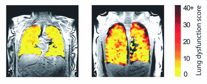
Photo caption: Lung function MRI showing problem areas (measurement levels of dysfunction) in lung transplant recipients.
Photo credit: Newcastle University, UK