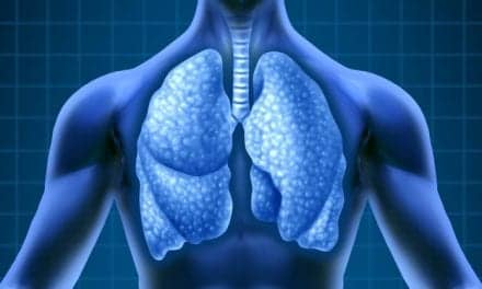
The challenge of deep particle deposition must be met with knowledge of aerosol fundamentals and the appropriate equipment.
Given the popularity of one aerosolized b-agonist, it might appear that contemporary aerosol therapy is exclusively concerned with bronchodilation. It is certainly true (and for good reason) that bronchodilators make up the lion’s share of drugs administered to patients via the aerosol route. They are, however, far from being the only agents that can be inhaled in particulate form. Barach et al1 first described the use of an aerosolized antibiotic for the treatment of a pulmonary infection as early as 1942. Since then, aerosol therapy using many different antimicrobial agents (such as gentamicin, carbenicillin, kanamycin, polymyxin, tobramycin, ribavirin, and pentamidine) has been reported, with varying degrees of success. Further, an even greater variety of nonantimicrobial agents have been aerosolized in recent years. This group has included various corticosteroids and other drugs such as amiloride, antiproteases, cyclosporine, heparin, insulin, morphine, prostacyclin, surfactant, and recombinant human DNase (rhDNase). Aerosolization of some of these drugs remains in the investigative stage at present, although inhaled insulin is nearly ready for commercial use and tobramycin and rhDNase are now commercial products (used primarily to treat cystic fibrosis). Other drugs will almost certainly become available for use as aerosols over the next few years.
One could argue that aerosolized bronchodilator administration is on autopilot. After all, it has evolved to become the epitome of routine therapy. User-friendly metered-dose inhalers (MDIs), dry-powder inhalers, and simple pneumatic small-volume nebulizers (SVNs) and air compressors have become ubiquitous. This type of aerosol therapy is being widely performed by RCPs, nurse practitioners, nurses, physicians, emergency medical personnel, school nurses, and physicians’ office assistants, not to mention patients and their families. This is good because the timely administration of aerosolized bronchodilators is a very significant factor in the successful treatment of asthma and related pulmonary conditions. The unfortunate aspect is the complacency that may have developed with respect to the technical requirements of aerosol therapy. Giving aerosol therapy looks too easy, but it may not always be as easy as it looks.
Arguably, providing aerosolized bronchodilator therapy requires little finesse and only a small amount of attention to detail. The nebulizer does not need any special attributes, and particle size distribution is not particularly crucial. For bronchodilator therapy, a nebulizer is not always required, inasmuch as MDI formulations of many bronchodilators are widely available. The site of aerosolized bronchodilator deposition may not be particularly important either, because b-agonists absorbed in upper airways can reach and stimulate receptor sites in lower airways through the bronchial circulation. Such is not necessarily the case, however, for other agents that require deeper pulmonary deposition for the treatment of endobronchial infection, for example. In the case of most of the experimental new drugs, the first (and, possibly, only) method of administration will be nebulization because MDI preparations are not unavailable. Therefore, it is important that practitioners be able to distinguish between the methods and devices routinely used to administer aerosolized bronchodilators and the more demanding techniques and devices used to administer aerosols that require deep deposition.
Fundamental Issues
One of the four fundamental issues in aerosol therapy is the dose-response relationship. It is traditional, in pharmacotherapy, to correlate the administered dose of a therapeutic agent with the response that its administration elicits. That is rarely possible for aerosolized drugs, however. The second fundamental issue is drug delivery. Unlike an oral tablet or intravenous injection, the dosage that is actually delivered to the patient may be significantly less than the dosage intended. This issue becomes even more complex when the site of drug delivery in the pulmonary system is considered. The third issue is deposition, a term widely misused and misunderstood. The fourth issue involves the size of the particles. Both MDIs and nebulizers have evolved from simple delivery devices into complex systems.2 As a result, many factors in addition to particle size also play important parts in the efficiency of those systems.
Dose Response
While the amount of medication placed in a nebulizer (or emitted by an MDI) is known, clinicians almost never know, in the typical clinical situation, how much of the drug is actually inhaled and retained by the patient. Ample investigation demonstrates that there is a very large and variable discrepancy between the nebulizer charge and the inhaled mass.3 Therefore, it is pointless to refer to what is placed in the nebulizer as the dose (although that convenient habit may be unbreakable). The assessment of bronchodilator response is relatively simple, and that response is nearly immediate.
The assessment of response to other drugs is less easy. A clinical practice guideline of the American Association for Respiratory Care4 states, “Although subjective responses and changes in mucociliary activity are important bronchodilator therapy effects, this guideline emphasizes the airway smooth muscle response that is primarily quantified through measurement of pulmonary function.” The guideline goes on to discuss various pulmonary function flow rate measures that can be used to evaluate bronchodilator response. The message is that aerosolized bronchodilator response can be readily and almost instantly assessed using physiologic measures such as peak flow; forced expiratory volume in 1 second; forced vital capacity; forced expiratory flow, midexpiratory phase; and simple clinical assessment (physical examination with chest auscultation). Bronchodilators have a very high toxicity threshold so that, in emergency situations, it is not unreasonable to administer repeated serial doses until an appropriate clinical response occurs or until some signs of toxicity appear. This approach is taken routinely, as evidenced by multiple-puff MDI protocols and continuous nebulization protocols for b-agonists.
In contrast, the assessment of other agents given by the aerosol route is extremely difficult and imprecise. With most drugs other than bronchodilators, there is a delayed response. For an inhaled antibiotic aerosol, for example, it may require days or weeks to determine whether any beneficial response has taken place and, if so, its degree. Complicating matters is the fact that the actual dose being delivered by a typical medication nebulizer is virtually unknown to the practitioner. It may be estimated in the aerosol laboratory using difficult and time-consuming techniques, but those tests can characterize only the effects of a specific set of variables (such as nebulizer brand, flow rate, volume of fill, drug concentration/dilution, patient tidal volume, respiratory rate, and duty cycle) on aerosol delivery. In clinical practice, there are so many permutations of these variables that the real dose is never known and the response cannot be directly correlated with the dose.
Drug Delivery
Because one cannot know with certainty what the delivered dose of an aerosolized drug actually is, other information that characterizes the performance of the aerosol device is extremely valuable. The term inhaled mass has been proposed by Smaldone3 to describe that fraction of the mass of drug placed in a nebulizer that would actually be inhaled by the patient. Inhaled mass can be determined on the test bench using a mechanical lung model, a simulated breathing pattern created by a ventilator, and filters that capture the nebulizer output that would have been inhaled. The influence of changing variables such as nebulizer fill volume, flow rate, breathing rate, tidal volume, and inspiratory time can be determined. Thus, inhaled mass describes the performance or efficiency of a delivery device under specified circumstances (see Figure 1, page 80). Nebulizers are relatively inefficient; as most practitioners routinely observe with practically all nebulizers, there is still liquid remaining in the device after it stops producing aerosol. That is the dead or residual volume, which is, to some extent, an indicator of inefficiency. Bench tests using pentamidine5 and saline6,7 with various nebulizers have shown that the type of nebulizer greatly influences the amount of drug that can be inhaled, and that some SVNs are more efficient than others.
Inhaled mass can also be determined in vivo using a combination of mass balance and filter collection techniques (with spontaneously breathing patients).8 If two different devices are tested in vivo under the same specified conditions, then inhaled mass provides a quantitative comparison. It permits the determination of the rate of drug delivery or output of a device and allows calculation of the total amount of drug delivered. Inhaled mass is expressed either as a percentage or as the absolute mass of drug inhaled. A single test is hardly illuminating, however. The real value of the inhaled mass assessment is that it allows comparison of two or more devices or allows techniques and conditions to be varied in order to assess their effects on nebulizer efficiency. This is important because, without some idea of the amount of drug leaving the nebulizer and being inhaled, it is not possible to distinguish between an inefficient nebulizer and abnormal patient physiology. Inhaled mass does not indicate how much of the inhaled drug was deposited or where it was deposited.
Deposition
A portion of the aerosol inhaled by a patient does not deposit in the airways or alveoli and is exhaled in the subsequent breath. The amount that does deposit is known as the deposition fraction. Obviously, the main goal of aerosol therapy is to increase the deposition fraction; the goal of deep aerosol therapy is to target the deposition to smaller peripheral airways and alveoli. The deposition fraction is dependent on three factors. The first is the patient’s pulmonary physiology; for example, a rapid, shallow breathing pattern and/or obstructive airway disease will reduce the deposition fraction. Second, the importance of the inhaled mass should not be underestimated. If the aerosol delivery device is inefficient, inhaled mass and deposition fraction will be reduced as a direct consequence.8 Third, the particle size distribution produced by the aerosol device must be conducive to impaction and sedimentation in the smaller airways. The customary generalization with respect to particle size is that larger particles tend to be deposited in upper airways; smaller particles tend to be deposited in smaller airways and, possibly, some alveoli; and the smallest particles tend to be exhaled.
Deposition fraction cannot be simulated on the test bench; it can be measured only in vivo. Measurement of deposition fraction usually involves conducting a deposition lung scan. A drug or solution in a nebulizer is tagged with a radioactive label and the patient’s lung fields are imaged using a gamma camera while the aerosol is inhaled. Assessment of deposition can be made by mapping various regions of interest. The radioactive counts in specific regions of interest are compared quantitatively to develop deposition ratios. For example, a high central-to-peripheral (C/P) ratio would indicate greater central deposition, while a lower ratio would indicate greater peripheral deposition. Thus, the site of deposition of an aerosol administered to target small airways could be evaluated through the use of the C/P ratio. The upper-to-lower lung and left-to-right deposition ratios may also be determined using the same technique, if required.
Particle Size Distribution
Clinical nebulizers are polydisperse devices; they create particles that span a wide range of dimension rather than a single size. Mass median aerodynamic diameter (MMAD) is used to describe a population of spheroid aerosol particles suspended in air or oxygen mixtures. MMAD is directly related both to diameter of the particles in the population and to the square root of the particle density. Generally, particle diameter has the greater effect on MMAD. When MMAD is measured, the resulting value indicates the median particle diameter. The geometric SD further describes the distribution around the median: a polydisperse aerosol has a geometric SD greater than 1.22, while a monodisperse aerosol has a geometric SD less than 1.22. As a term, MMAD is used by aerosol scientists to describe particle size distribution because individual particle sizes vary widely in a polydisperse aerosol.
The term may also be used as an indicator of the performance of devices, but a stated MMAD represents the median–not necessarily the average–particle size, and it certainly does not represent the size of the majority of particles. Even more confusing are statements that a particular device produces an MMAD of, for example, 3 to 5 mm. The MMAD can be only a single value, not a range.
Clinical Implications
Some particles are too small to be deposited and will be exhaled, while others are too large to escape the nebulizer in the first place. Some of the particles that do leave the nebulizer may be so large as to be deposited in the oropharynx or upper airways, while others may be the optimum size for deposition at the target site. This correlates with the concept of a particle size distribution, rather than a single dimension.
What is the optimum particle size? Many textbooks state that particles of 3 to 5 mm have the greatest likelihood of being deposited in the human tracheobronchial system. While this may be true of aerosolized bronchodilator therapy in adults, it may not be true of deep aerosol therapy targeting the terminal bronchioles and alveoli (or even for general aerosol therapy in small children and infants). Smaller particles will be necessary for deep aerosol therapy; under some circumstances, an MMAD of 1 æm may be far more appropriate than an MMAD of 3æm.
Summary
Another often-overlooked aspect of aerosol therapy is that as the particles become smaller, the mass of drug they can carry diminishes. The volume of a sphere is the following:
Volume of a Sphere = 4πr3/3 (where r=radius of the particle)
For a spherical particle with a diameter of 4 mm, the effect of cubing the sphere’s radius on the mass of drug can be expressed as 2x2x2=8 units of mass. If the particle size is reduced from 4 mm to 2 mm, then the cubed radius (1x1x1) produces only one unit of mass. Thus, if the particle is too small, it can not carry very much medication, and compensation may be have to be provided in the form of a greater aerosol output (increased inhaled mass). On the other hand, if the particle size is increased from 4 mm to 8 mm, then 4x4x4=64 units of mass. Although a great deal of drug is carried in these large particles, their chance of reaching the smaller airways is practically nil.
Conclusion
Unlike bronchodilator therapy, deep particle deposition is a challenge that requires exceptionally capable equipment and the knowledge to use it effectively. The aerosol delivery system has a profound effect on the delivery of an efficacious dose of antibiotics (and other nonbronchodilator drugs) to small airways and alveoli. For those drugs for which deep deposition is needed, the practitioner must select and use a delivery system that is more efficient and precise than that required for bronchodilator treatment. An understanding of the fundamental concepts of dose response, drug delivery, deposition, and particle size distribution is a clinical imperative in the making of that selection.
Michael McPeck, RRT, is director, respiratory care and biomedical engineering, University Medical Center, State University of New York, Stony Brook.
References
1. Barach AL, Molomut M, Soroka M. Inhalation of promin in experimental tuberculosis. American Review of Tuberculosis. 1942;46:268-276.
2. McPeck M. Aerosol research: how should we skin this cat? [editorial]. Respiratory Care. 1994;39:1155-1156.
3. Smaldone GC. Drug delivery via aerosol systems: concept of “aerosol inhaled.” Journal of Aerosol Medicine. 1991;4:229-235.
4. AARC Clinical Practice Guideline ARBD: assessing response to bronchodilator therapy at point of care. Respiratory Care. 1995;40:1300-1307.
5. Smaldone GC, Perry RJ, Deutsch DG. Characteristics of nebulizers used in the treatment of AIDS-related Pneumocystis carinii pneumonia. Journal of Aerosol Medicine. 1988;1:113-126.
6. Hess D, Fisher D, Williams P, Pooler S, Kacmarek RM. Medication nebulizer performance: effects of diluent volume, nebulizer flow, and nebulizer brand. Chest. 1996;110:498-505.
7. Tandon R, McPeck M, Smaldone GC. Measuring nebulizer output: aerosol production vs gravimetric analysis. Chest. 1997;111:1361-1365.
8. Smaldone GC, Fuhrer J, Steigbigel RT, McPeck M. Factors determining pulmonary deposition of aerosolized pentamidine in patients with human immunodeficiency virus infection. Am Rev Respir Dis. 1990;143:727-737.


