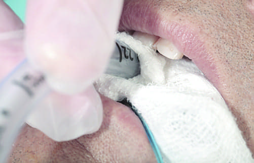
With rumors that ventilator-associated events could be added to the CMS never event list (which leads to no reimbursement), medical institutions have even more vested interest in preventing these conditions.
It’s been nearly three years since new definitions for ventilator-associated events (VAEs) were released by the Centers for Disease Control and Prevention’s National Health Safety Network (NHSN). Since then, there have been some small bumps—several modifications have been made to the definitions in response to user issues—but in general, the healthcare world has continued moving forward much the same.
“The criteria for identifying VAEs does not involve changes in ventilator management strategies that are already known,” said Paul Garbarini, MS, RRT, Vice President Product Research and Development for Hamilton Medical Inc, Reno, Nev.
What the new definitions have done, however, is provided quantitative and qualitative criteria through surveillance that the medical community can use to research the incidence and prevention of VAEs—outside of VAP. New studies have begun to focus on establishing the validity of management strategies, and new technologies are helping to implement them more easily. Moving forward, the new definitions may have more impact than when they first were implemented.
The investment made in new research and technology is well worth the return in terms of money saved and, more importantly, improved outcomes. It is estimated that more than 300,000 patients receive mechanical ventilation in the United States each year.1 These patients are at risk for a number of the newly defined VAEs. One recent study found that among 3,028 mechanically ventilated patients, 77% had at least one ventilator-associated condition, and 29% had one infection-related complication episode.2
Mortality rates are also grim. Estimates in patients with acute lung injury on mechanical ventilation range from 24% in patients aged 15 to 19 years to 60% in those 85 years and older.1
Nor do these poor outcomes come cheaply. Scott calculates the range of the average cost attributable to ventilator-associated pneumonia (VAP), the most studied VAE, between $19,000 and $29,000 in his publication on the direct medical costs of healthcare-associated infections in US hospitals and the benefits of prevention.3 The Institute for Healthcare Improvement cites an even higher figure of $40,000 in additional costs for a typical hospital admission with VAP.4
With rumors that VAEs could be added to the “never” event list, which leads to no reimbursement, medical institutions have even more vested interest in preventing these conditions. Best practice protocols can help, and using the clearer definitions and validated studies, hospitals are revising their management programs to incorporate new information and new technology.
What’s a VAE?
In developing the surveillance definition algorithm, the NHSN sought to use “objective, streamlined and potentially automatable criteria” to identify the broad range of conditions and complications seen in mechanically ventilated adult patients.1 The standards were designed to track episodes of sustained respiratory deterioration in mechanically ventilated patients after a period of stability or improvement.
The objective points of comparison include deterioration in respiratory status after a period of stability or improvement on the ventilator, evidence of infection or inflammation and laboratory evidence of respiratory infection. Using these criteria, medical institutions can track and report on VAEs as a whole, and not just VAP.
Prior to the definition change, VAP was the primary VAE tracked by the healthcare community. The new VAE algorithm breaks VAEs into three definition tiers: ventilator-associated conditions (VACs), infection-related ventilator-associated complications (IVACs) and possible VAP (PVAP).
The majority of these are typically attributed to one of four causes: pneumonia, pulmonary edema, atelectasis and acute respiratory distress syndrome (ARDS).1,5 Garbarini suggested barotrauma and ventilator-induced lung injury (VILI), which leads to ARDS, could also be added to the list.
Ventilator Management Strategies
Efforts to prevent VAEs, therefore, tend to focus on the prevention of these conditions, with many targeting VAP. The most common suggestion for avoiding any of them, but particularly VAP, is to minimize ventilation time and, if possible, avoid invasive mechanical ventilation altogether. This is clearly not always possible.
When that is the case, other potential strategies include preventing aspiration, minimizing sedation, performing paired daily spontaneous awakening and breathing trials, encouraging early exercise and mobility, using low tidal volume ventilation, incorporating conservative fluid management, implementing conservative blood transfusion thresholds, reducing gastric colonization and decreasing the risk of contamination of respiratory equipment.
Interventional studies published to this point have collected evidence that shows minimizing sedation, paired daily spontaneous awakening and breathing trials and conservative fluid management can reduce VAE rates and improve patient-centered outcomes.5 Klompas suggests that new studies should look at other interventions, additional modifiable VAE risk factors and prevention bundles that improve outcomes even more when used together than alone.5
In the meantime, hospitals continue to implement VAE management strategies that incorporate as many of the methods that they are capable of implementing. These will likely be expanded and refined much as the definitions were, allowing RTs to develop more effective care protocols and choose more supportive technologies to aid their efforts toward the prevention of VAEs.
Keep Noninvasive an Option
There is already equipment available to assist in many of the existing efforts in use today. Ventilation technology can even help with the number one goal of minimizing the amount of time spent on a ventilator. Fewer ventilated days translates to less risk for a VAE, particularly VAP.
Garbarini notes that all Hamilton Medical ventilators provide invasive as well as noninvasive ventilation support (NIV). Many other manufacturers, such as Maquet and Covidien (now part of Medtronic), have similar offerings. Users can switch between modes without having to switch equipment or rooms and avoiding a potential negative impact on the outcome.
Some ventilator models are designed for portability so they may also be used in transport and to aid with early mobility efforts. Lightweight and battery-powered, they permit greater flexibility in a variety of settings. Hamilton has the Hamilton-T1 portable ventilator, which offers the performance of a fully featured ICU ventilator with more than nine hours of battery time. The portable Flight 60 by Maquet (Wayne, NJ) offers invasive and noninvasive ventilation modes with advanced monitoring capabilities, such as plateau pressure and static compliance, that can run up to 12 hours using a hot swappable battery. And Medtronic’s ruggedly designed Newport HT70 Plus Ventilator can run up to 10 hours with advanced features such as Alarm Quickset, custom presets and custom backup ventilation.
Wean STAT
When a patient does require invasive ventilation, having noninvasive ventilation on the monitor can assist with weaning efforts. Garbarini points to a 2013 study by Burns, et al, where noninvasive weaning was found to reduce the rates of death and pneumonia without increasing the risk of weaning failure or reintubation.6 The researchers also found mortality benefits were significantly greater in patients with COPD in a subgroup analysis.6
The ability of patients to be successfully weaned, with or without noninvasive ventilation, however, is often underestimated by clinicians. Ventilator equipment can assist, helping the RT to assess and identify a patient’s readiness to wean, conduct spontaneous breathing trials (SBTs), prevent ventilator-induced diaphragm dysfunction and promote early mobility.
With most advanced equipment, RTs can check on a patient’s status quickly using dashboard systems. Hamilton Medical ventilators feature the Vent Status panel, which displays the dependency of the patient on the ventilator, tracks six key parameters against weaning criteria, and indicates when the patient is ready for extubation. Floating indicators and colored lights help deliver instant visual information at a glance.
It displays data generated by the company’s Intellivent-ASV technology, which uses patient data and targets and is currently only available outside of the US, such as those for EtCO2 and SpO2, to help RTs automatically select ventilator settings, manage the transition from passive to spontaneous breathing ventilation and provide an automated weaning protocol.
Studies have shown that enhanced performance of paired, daily SATs (spontaneous awakening trials) and SBTs is associated with lower VAE rates.5 Klompas found that within collaborative units (though not surveillance units), significant increases in SATs, SBTs and percentage of SBTs performed without sedation were mirrored by significant decreases in the duration of mechanical ventilation and hospital length-of-stay.5
To prevent autopeep, enhance synchrony, and decrease ventilator-dependence, Hamilton’s ASV mode will automatically transition patients from full to partial support when they begin to breathe spontaneously. Default target ranges that can be manually modified are set when the RT selects the patient’s clinical condition. Other parameters, such as the tidal volume and respiratory rate, are determined and adjusted by the algorithm according to criteria.
The system also works with an optional weaning protocol, Quick Wean, that includes the ability to automatically conduct controlled SBTs as well as an operator-configurable weaning protocol. It decreases pressure support progressively and screens for the readiness-to-wean criteria.
Minimize Volutrauma
The parameters and principles employed to automate weaning can also be applied to minimize other complications as well. To prevent volutrauma due to lung overdistention, the Hamilton Intellivent-ASV mode will maintain an operator-set minute volume and automatically determine an optimum tidal volume-respiratory rate combination based on the minimal work of breathing principle described by Otis in 1954.
According to Garbarini, targets are based on research showing that low tidal volumes can reduce mortality in patients with ARDS. He cites a 2012 study by Needham et al that found that despite the evidence supporting the positive effect of this strategy, as few as 40% of ARDS patients actually receive these protective tidal volumes.7 The Intellivent-ASV values are set to maintain tidal volumes that match goals suggested by the ARDSnetwork: between 4 to 8 mL/kg/IBW with plateau pressure below 30 cm H2O.
The system uses a proximal flow sensor to measure the patient’s respiratory mechanics breath-by-breath. The algorithm is adjusted to match all stages, from passive ventilation to spontaneous breathing to weaning.
Some patients, such as the morbidly obese, may be able to withstand higher pressures. This is typically determined via transpulmonary pressure calculations using esophageal pressure, according to Garbarini. Transpulmonary pressure monitoring is available as a standard feature with the Hamilton-G5 and Hamilton-S1 ventilators.
Plus, the company has also developed the Protective Ventilation Tool (P/V Tool) that provides a simple, repeatable maneuver to record a quasi-static pressure/volume curve and can be used in conjunction with esophageal pressure monitoring to identify and avoid volutrauma and/or overdistension. It employs a patented pressure-ramping technique that produces different curves that can be used to analyze the recruitment strategy.
Avoid Atelectrauma
Analysis of these curves provides additional information that can be used to prevent other forms of VAE. Determining P/V loop inflection points and/or hysteresis, which can be done rapidly with the technologies like Hamilton’s P/V tool, can help RTs to identify optimal PEEP settings. Individualizing PEEP titration based on a quasi-static pressure/volume loop is thought to help prevent atelectrauma.
Research on atelectrauma points to the repetitive opening and closing of the airways and lungs during mechanical ventilation as a cause. Gattinoni et al suggest “that stress at rupture is only rarely reached and that high-tidal volume induces VILI by augmenting the pressure heterogeneity at the interface between open and constantly closed units.”8 The study hypothesizes that VILI occurs only when a given threshold is exceeded and that, below this limit, mechanical ventilation is likely to be safe.8 The P/V Tool, and others like it, can therefore help to identify the limits, not only for appropriate ventilation settings but also recruitment maneuvers.
PEEP alone may not be sufficient to recruit significant numbers of collapsed alveoli. For some patients, high threshold opening pressures may be required to reopen them, but these same pressures could also reinjure a nonrecruitable lung. Assessment of hysteresis with Hamilton’s unique pressure-ramping technique correlates with lung recruitablity, provides a measurement of recruited lung volume and allows the RT to configure individualized automated lung recruitment maneuvers.
Cuff VAP
Of course, the clinician can get all of the settings right, and the patient can still contract a ventilator-associated infection. As one of the few measured VAEs prior to 2013, much research has been done in this area. In addition to minimizing the time spent on ventilation and routine hygiene procedures during, minimizing contamination via microaspiration is also a major target for prevention.
“Microaspiration of secretions past the ETT cuff is a primary causative risk factor for VAP. So RTs want to adhere to recommendations to maintain cuff pressures at 20 to 30 cm H2O,” says Garbarini.
However, to stay within this optimal range can require up to eight adjustments per day. Systems that automate this protocol can help to more consistently maintain the appropriate cuff setting without requiring time or staff resources. Hamilton offers IntelliCuff, a cuff pressure controller that automatically adjust cuffed endotracheal and tracheostomy tubes to match the clinician-set parameter.
Other manufacturers have developed their own unique technologies and systems to aid RTs in the management of ventilated patients. With options for invasive and noninvasive ventilation, portability, breath monitoring and control, care protocols and weaning, technology can help RTs prevent VAEs and improve the outcomes of their patients. Whatever reimbursement issues exist, clinicians already aim to have VAEs on their “never” lists. The new definitions are enabling them to create better protocols and discover build better tools to get patients breathing on their own. RT
Renee Diiulio is a contributing writer to RT. For further information, contact [email protected].
References
-
Ventilator-associated event. Device Associated Module. Centers for Disease Control and Prevention National Health Safety Network. January 2015. Available at http://www.cdc.gov/nhsn/PDFs/vae/Draft-Ventilator-Associate-Event-Protocol_v6.pdf. Accessed on October 12, 2015.
-
Bouadma L, Sonneville R, Garrouste-Orgeas M, et al. Ventilator-associated events: Prevalence, Outcome, and Relationship Ventilator-Associated Pneumonia. Crit Care Med. 2015 Sep;43(9):1798-806.
-
Scott RD II. The direct medical costs of healthcare-associated infections in U.S. hospitals and the benefits of prevention. Division of Healthcare Quality Promotion National Center for Preparedness, Detection, and Control of Infectious Diseases, Coordinating Center for Infectious Diseases, and Centers for Disease Control and Prevention. March 2009.
-
Institute for Healthcare Improvement. How-to guide: Prevent Ventilator-associated pneumonia. 2012. Available at http://www.ihi.org/resources/Pages/Tools/HowtoGuidePreventVAP.aspx. Accessed on October 12, 2015.
-
Klompas M. Potential strategies to prevent ventilator-associated events. Am J Respir Crit Care Med. September 23, 2015. Available at ww.atsjournals.org/doi/abs/10.1164/rccm.201506-1161CI#.VhwTfqR9YeN. Access on October 11, 2015.
-
Burns K, Meade M, Premji A, and Adhikari N. Noninvasive ventilation as a weaning strategy for mechanical ventilation in adults with respiratory failure: a Cochrane systematic review. Canadian Medical Association Journal. 2013;186(3):E112-E122.
-
Needham D, Colantuoni E, Mendez-Tellez P, et al. Lung protective mechanical ventilation and two year survival in patients with acute lung injury: prospective cohort study. BMJ. 2012;344:e2124-e2124.
-
Gattinoni L, Protti A, Caironi P, and Carlesso E. Ventilator-induced lung injury: the anatomical and physiological framework. Crit Care Med. 2010 Oct;38 (10 Suppl):S539-48.


http://www.mercurymed.com/catalog2/index.php?manufacturer=44