Endotracheal, nasotracheal, and tracheostomy tubes can provide a high level of support for critically ill patients—if effective management of these artificial airways is maintained.
By Bill Pruitt, MBA, RRT, CPFT, FAARC
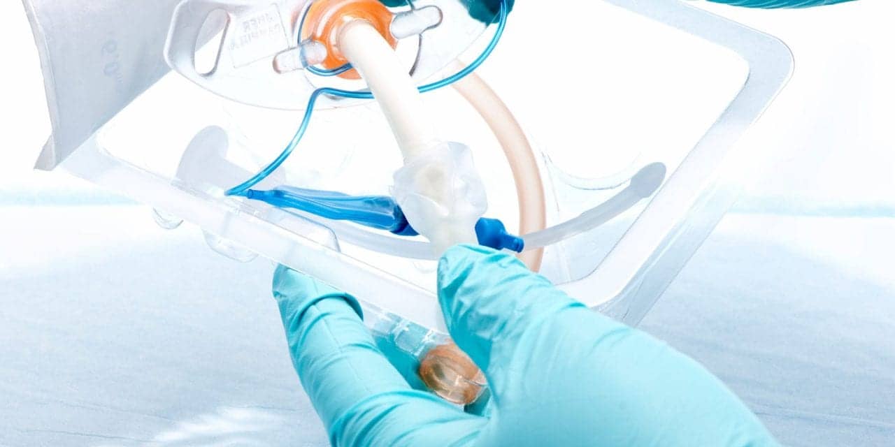
Sometimes patients need to have the highest level of support in maintaining oxygenation, ventilation, or in protecting their natural airway. By placing an artificial airway through inserting an endotracheal tube (ETT), nasotracheal tube (NTT) or placing a tracheostomy tube by surgery or by percutaneous dilation, this high level of support can be established. Once in place it is important to secure the airway and use the care to avoid complications related to these devices.
This article will explore these intubation issues and give some practical, proven tips to provide the best care in airway management.
Indications and Procedures
An artificial airway—such as endotracheal, nasotracheal, or tracheostomy tubes—are needed for several cardiopulmonary-related conditions. These issues include:
- Obstruction of the airway by excess mucus, inflammation, or external pressure (ie, a tumor causing airway collapse).
- Cardiac or respiratory arrest where the cardiac or pulmonary function ceases.
- Trauma or some injury to the chest, neck, or abdomen that compromises the airway or diaphragm.
- Loss of consciousness bringing about inability to protect the airway.
- General anesthesia for surgery or deep sedation for procedures (resulting in no respiratory effort or severely compromised breathing).
- Severe sleep apnea that cannot be managed by other approaches.
- Increase risk to the airway due to aspiration, lack of gag or cough reflex, excessive bleeding.1
Artificial airways may be needed in an emergency situation (ie, cardiopulmonary arrest or as the result of trauma or burns) or may be placed electively (prior to a procedure, such as receiving anesthesia for surgery) or impending cardiopulmonary failure with deteriorating status of the patient.
The most common placement of an artificial airway is oral intubation to insert an ETT. In this procedure, an endotracheal tube is passed through the mouth, (oral cavity) and pharynx (including larynx, and vocal cords) into the trachea using visualization of passage by laryngoscope (often using video like the GlideScope or CMAC to improve visualization). Other aids in intubation include a bougie, lighted stylet, or optical stylet. ETT placement can also be accomplished by passing the tube over a bronchoscope.
A nasotracheal tube (NTT) passes through the nares, through the naso, oro, and laryngopharynx into the trachea, and can be done as a “blind” procedure (no visualization) or aided by visualization with a laryngoscope or bronchoscope. Nasotracheal intubation is rare and is used in patients who would have difficulty with the oral placement (i.e. burn patients, those with trauma to the jaw, or those undergoing surgical procedures where an oral ETT would interfere with the surgical field). NTTs have higher risks of complication (increase the risk of sinusitis, bleeding, pain) and often require a smaller size tube compared to using the oral approach (smaller tubes result in higher resistance to airflow, increasing work of breathing and making suctioning more difficult).2
Tracheostomy tubes are placed by a surgical procedure in an operating room (OR) or outside the OR, using a percutaneous approach (accomplished by dilation of a small opening in the trachea and aided by bronchoscopy to insert the tracheostomy tube).
A tracheostomy is most commonly used in cases of prolonged mechanical ventilation where an oral ETT has been used initially and it is anticipated that ventilatory support will continue. Other indications for a tracheostomy include neuromuscular disorders, or other conditions that are not responsive to therapy (ie severe obstructive sleep apnea, subglottic stenosis, vocal cord paralysis).3
Compared to the oral ETT, tracheostomy offers several advantages in areas such reduced airflow resistance in less work of breathing, allowing for speech and swallowing, easier suctioning, increased patient comfort, better oral care and oral hygiene, and a more stable airway (especially during ambulation or transport of the patient).3
Securing Endotracheal, Nasotracheal, Tracheostomy Tubes
ETTs have been secured for years by use of adhesive tape or twill tape, but the trend now has shifted to using a commercial ETT holder. Migration of the ETT in the airway (either deeper or more shallow) can lead to unilateral ventilation or accidental extubation depending on which direction the tube moves. A migration of the ETT can also increase the risk of a ventilator-associated event (VAE) by under or over-ventilating the lung(s) or ventilator-associated pneumonia (VAP) due to increased aspiration.
Adhesive tape has the advantages of low cost and good “holding power” to prevent accidental extubation but presents difficulties in moving the tube from one side to the other as the tapes has to be removed and a new taping procedure performed once the move is finished. Other issues with tape include losing the adhesive grip in the face of copious secretions, and the learning curve needed to become proficient and efficient in applying the tape in an acceptable, effective manner. There are also problems with pressure necrosis at the side/corners of the mouth, especially if some of the skin at the corners of the mouth gets caught against the ETT.
Commercial ETT holders offer the advantages of being able to adjust the ETT position, both in depth and in moving from one side of the mouth to the other with greater ease compared to adhesive tape or twill. Commercial ETT holders may also include a bite block that prevents the patient from biting down on the ETT. Disadvantages the commercial ETT holders include increased cost, increased risk of skin issues leading to pressure sores and necrosis, and kinking of the ETT.4
There are many commercial ETT holders on the market including AnchorFast ET Tube Holder (Hollister, Libertyville, IL), Cooper Surgical Neo-fit (Cooper Surgical, Trumbull, CT), Smiths Medical Portex ETTube Holder (Smiths Medical, Dublin, OH), Neotech Neo Bar (Neotech, Valencia, CA), Ambu ETT Holder (Ambu, Ballerup, Denmark), and the Laerdal Thomas Tube Holder (Laerdal, Wappingers Falls, NY). Published studies have compared various approaches to securing the ETT but have not found consensus as to which approach is the best overall.5
Cuff Management
Cuffed artificial airways (both ETT and tracheostomy tubes) are useful in preventing aspiration of secretions and allow for the use of positive pressure to ventilate the patient (or apply CPAP, BiPAP, and PEEP). In newborns and young patients uncuffed artificial airways may used if there is minimal to no leak around the tube. Cuffed ETTs and tracheostomy tubes have been designed to reduce the amount of pressure exerted on the tracheal wall. Years ago tubes utilized cuffs that were more bulbous in shape and were classified as low volume, high pressure cuffs, but changes in design have made cuffs longer or more tear-drop shaped and have changed to be classified as high volume, low pressure cuffs. Use of cuffed airways calls for careful monitoring and adjustment if the cuff does not seal well (under-inflated) or is over-inflated. Lack of a good seal will lead to loss of pressure/volume in positive pressure applications, increased risk of aspiration, and increased risk of accidental extubation. Over-inflated cuffs can lead to excessive pressure on the lateral tracheal walls, blocked circulation through the capillary bed, and infarction resulting in tracheomalacia (collapse of the airway walls due to weakness). The general consensus is maintain cuff pressures between 20-30 cmH2O. (However, at times it is not possible to keep a good seal during positive pressure ventilation unless the cuff pressure is higher than 30 cmH2O).5
Various techniques have been developed to try and obtain the best cuff pressure (including the minimal leak technique and the minimal occluding volume technique) and many institutions use cuff manometers to facilitate checking and adjusting cuff pressure. There are many commercial devices for checking and altering cuff pressures including the Posey Cufflator and the Rusch Endotest Cuff Pressure Manometer. Devices that continuously measure and maintain cuff pressures include the Puritan Bennett Cuff Pressure Manager, the Hamilton Medical Intellicuff, The ARM Medical Pyton, and the Outcome Solutions Cuff Sentry. Disposable devices include the Medline/Hospitech AG Cuffill and the Ambu Disposable Pressure Manometer. There are several studies that have evaluated issues with different devices related to accuracy, possible loss of pressure that may occur in connection and disconnection of the device to the pilot balloon, and other aspects of these devices.6-8
Suctioning
Suctioning the airway is done to remove secretions in order to reduce resistance, prevent blocked airways, mucus plugs, and atelectasis, and to reduce the incidence of nosocomial infection due to retained secretions. Suctioning can be performed by single use, disposable suction catheters which require the patient circuit to be disconnected to perform the procedure (classified as open suctioning system) or by suction catheters that are multiple use, enclosed inside a collapsible plastic bag, and left in place as part of the patient circuit (classified as closed suction system). In 2022, the American Association for Respiratory Care (AARC) published an excellent resource in their clinical practice guideline on suctioning that covers these two systems as well as many other aspects of the procedure including depth of suctioning, suction pressures, use of saline lavage, duration of suctioning, suction catheter sizes, etc.9
Subglottic suctioning included in the design of ETTs is another aspect of airway care that has been evaluated for a means to reduce ventilator associated pneumonia (VAP) and impact the patient’s overall stay in the hospital. Tubes with this design have a small opening above the cuff that connects to a channel running up the inside of the ETT and ending in a port. The port is connected to a low pressure suction setup to remove secretions that pool in the airway above the cuff. The suctioning can be continuous or intermittent.
Use of subglottic suctioning has been shown to reduce VAP, shorten length of time on mechanical ventilation, and shorten length of stay in ICU. The Agency for Healthcare Research and Quality (AHRQ) recommends using a strategy to identify high-risk areas of the hospital where aspiration of subglottic secretions may be more common and use Subglottic Secretion Drainage Endotracheal Tubes (SSD-ETTs) instead of standard ETTs in these areas.10
Infection Control
Artificial airways provide an open route between outside the body and inside the airways, bypassing the natural barriers found in the undisturbed airway (such as filtering and humidification in the nasal passages, the mucociliary escalator for removing foreign materials from the airways, cough reflex, etc). This situation increases the risk of infection and VAP.
Usual, customary infection control practices must be utilized when dealing with an artificial airway, including handwashing, use of personal protective equipment, proper use of disposable single patient use circuits and circuit filters, cleaning and disinfection of devices and machines, etc.
Use of a comprehensive approach to airway care (“ventilator bundle”) have been recommended to reduce infections, VAP, and ventilator-associated events (VAE).
Elements of various ventilator bundles utilized to reduce incidence of VAP include:11
- Keep the head of the bed elevated to 30-45 degrees;
- Avoid use of nasogastric tubes, limit the use of orogastric tubes;
- Provide oral care with chlorhexidine;
- Place “tonsil tip” suction devices in a protective cover between uses;
- Utilize spontaneous breathing trials and daily sedation interruption;
- Provide deep vein thrombosis and stress ulcer prophylaxis;
- Use ETTs with subglottic suction;
- Use silver-coated ETTs, ETTs with tapered cuff design, or ETTs made of polyurethane material;
- Use special catheters to remove biofilm from ETT lumen;
- Use automated monitoring and control of ETT cuff pressures;
- Replace ventilator circuits weekly or when visibly soiled;
- Use closed suction catheter system and replace using the same strategy as ventilator circuits;
- Use heat-moisture exchangers when appropriate;
- Use protective covers for self-inflating resuscitation bags; and
- Perform handwashing before and after contact with the ventilator or ventilator circuit.
Conclusion
Artificial airways are needed to protect the airway, provide a means for suctioning, and provide a secure and safe means to provide positive pressure ventilation. Airways can be established by several different approaches and must be secured in a safe, effective manner to avoid loss of the airway or causing some harm to the patient through pressure sores, increased risk of aspiration, etc. Clearing the airways of secretions by suctioning is often necessary but needs to be done with care using the most appropriate means. Ventilator bundles are very useful in providing steps to take to avoid VAP. Research in many airway-related areas is ongoing. Respiratory therapists need to stay up-to-date in the latest proven methods for managing artificial airways in order to provide the best, evidence-based care.
RT
Bill Pruitt, MBA, RRT, CPFT, FAARC, is a writer, lecturer, and consultant. Bill has over 40 years of experience in respiratory care in a wide variety of settings and has over 20 years teaching at the University of South Alabama in Cardiorespiratory Care. Now retired from teaching, Bill continues to provide guest lectures, participates in podcasts, and writes professionally. For more info, contact [email protected].
References
- Cleveland Clinic website on intubation: www.myclevelandclinic.org/health/articles/22160-intubation.
- Yamamoto R, et al. Nasal intubation for trauma patients and increased in-hospital mortality. European Journal of Trauma and Emergency Surgery. 2022 Aug;48(4):2795-802.
- From UpToDate website on Tracheostomy: https://www.uptodate.com/contents/tracheostomy-rationale-indications-and-contraindications.
- Branson RD, et al. Management of the Artificial Airway Discussion. Respiratory Care. 2014 Jun 1;59(6):974-90.
- Dexter AM, Scott JB. Airway management and ventilator-associated events. Respiratory Care. 2019 Aug 1;64(8):986-93.
- Asai S, et al. Decrease in cuff pressure during the measurement procedure: an experimental study. J of Intensive Care. 2014 Dec;2(1):1-5.
- Rackley CR. Monitoring during mechanical ventilation. Respiratory Care. 2020 Jun 1;65(6):832-46.
- Blanch PB. Laboratory evaluation of 4 brands of endotracheal tube cuff inflator. Respiratory care. 2004 Feb 1;49(2):166-73.
- Blakeman TC, Scott JB, Yoder MA, Capellari E, Strickland SL. AARC clinical practice guidelines: artificial airway suctioning. Respiratory Care. 2022 Feb 1;67(2):258-71.
- From the Agency for Healthcare Research and Quality (AHRQ) website on “The Benefits of Subglottic Secretion Drainage Endotracheal Tubes” 2017: https://www.ahrq.gov/hai/tools/mvp/modules/technical/subglottic-fac-guide.html. Accessed March 2023.
- Kallet RH. Ventilator bundles in transition: from prevention of ventilator-associated pneumonia to prevention of ventilator-associated events. Respiratory Care. 2019 Aug 1;64(8):994-1006.


