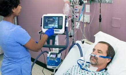
The FDA issued 510(k) clearance for GE Healthcare’s artificial intelligence (AI) algorithm to help clinicians assess endotracheal tube (ETT) placements. The AI solution is one of five included in GE Healthcare’s Critical Care Suite 2.0, an industry-first collection of AI algorithms embedded on a mobile x-ray device for automated measurements, case prioritization and quality control.
GE has distributed this solution under the FDA COVID-19 imaging guidance since November 2020, citing its potential to impact clinicians who were experiencing an influx of critically ill COVID-19 patients who required ventilation. Research shows that up to 25% of patients intubated outside of the operating room have misplaced ETTs on chest x-rays, which can lead to severe complications for patients, including hyperinflation, pneumothorax, cardiac arrest and death. Moreover, as COVID-19 cases climb, with more than 250 million confirmed worldwide, anywhere from 5-15 percent require intensive care surveillance and intubation for ventilatory support. Over the past year, 200 hospitals have deployed the technology to assist with ETT placements. The FDA has now granted 510(k) clearance for this solution allowing its continued sale outside of the public health emergency.
“At GE Healthcare, we saw the potential role of Critical Care Suite 2.0 in helping hospitals manage the crisis caused by the number of patients who needed ETT placements during the pandemic, requiring accelerated innovation, and we quickly worked with the FDA to make the solution available to clinicians. We are pleased to now have the FDA’s clearance for this important solution,” said Jan Makela, President and CEO, Imaging at GE Healthcare. “The pandemic has proven what we already knew – that data, AI and connectivity are central to helping front line clinicians deliver intelligently efficient care. GE Healthcare is not only accelerating the development and access of new tools to help hospital staff keep up with demand, but also leading the way on COVID-era advancements that will transform the industry and have a long-lasting impact after the pandemic.”
Up to 45% of ICU patients, including severe COVID-19 cases, receive ETT intubation for ventilation. While proper ETT placement can be difficult, Critical Care Suite 2.0 uses AI to automatically detect ETTs in chest x-ray images and provides an accurate and automated measurement of ETT positioning to clinicians within seconds of image acquisition, displaying the results on the monitor of the x-ray system. The AI generated measurements – along with an image overlay – are then made accessible in picture archiving and communication systems (PACS). The average error is less than 1.0mm when calculating the endotracheal tube tip-to-carina vertical distance. With these measurements, clinicians can determine if the ETT is placed correctly or if additional attention is required for proper placement.
Improper positioning of the ETT during intubation can lead to various complications, including a pneumothorax, a type of collapsed lung. While the chest x-ray images of a suspected pneumothorax patient are often marked “STAT,” they can sit waiting for up to eight hours for a radiologist’s review. However, when a patient is scanned on a device with Critical Care Suite 2.0, the system automatically analyzes images and sends an alert for cases with a suspected pneumothorax – along with the original chest x-ray – to the radiologist for review in PACS. The technologist also receives a subsequent on-device notification to provide awareness of the prioritized cases.
“Seconds and minutes matter when dealing with a collapsed lung or assessing endotracheal tube positioning in a critically ill patient,” explains Dr. Amit Gupta, Modality Director of Diagnostic Radiography at University Hospital Cleveland Medical Center and Assistant Professor of Radiology at Case Western Reserve University, Cleveland. “In several COVID-19 patient cases, the pneumothorax AI algorithm has proved prophetic – accurately identifying pneumothoraces/barotrauma in intubated COVID-19 patients, flagging them to radiologist and radiology residents, and enabling expedited patient treatment. Altogether, this technology is a game changer, helping us operate more efficiently as a practice, without compromising diagnostic precision.
To make the AI suite more accessible, Critical Care Suite 2.0 is embedded on a mobile x-ray device – offering hospitals an opportunity to try AI without making investments into additional IT infrastructure, security assessments or cybersecurity precautions for routing images offsite.
Furthermore, the on-device AI offers several benefits to radiologists and technologists:
- ETT positioning and critical findings: GE Healthcare’s algorithms are a fast and reliable way to ensure AI results are generated within seconds of image acquisition, without any dependency on connectivity or transfer speeds to produce the AI results.
- Eliminating processing delays: AI results are then sent to the radiologist while the device sends the original diagnostic image, ensuring no additional processing delay.
- Ensuring quality: The AI suite also includes several quality-focused AI algorithms to analyze and flag protocol and field of view errors as well as auto rotate the images on-device. By automatically running these quality checks on-device, it integrates them into the technologist’s standard workflow and enables technologist actions – such as rejections or reprocessing – to occur at the patient’s bedside and before the images are sent to PACS.
GE Healthcare worked with multiple academic research partners including UC San Francisco and the University Hospitals Cleveland Medical Center to develop the Critical Care Suite using GE Healthcare’s Edison platform, which helps deploy AI algorithms quickly and securely. Critical Care Suite 2.0 is available on the company’s AMX 240 and AMX Navigate mobile x-ray systems.