Northwestern University researchers have developed soft, miniaturized wearable devices that continuously and wirelessly monitor sounds inside patients’ bodies, such as air moving in and out of the lungs, offering new insights into lung health and other regions of the body.
The study is published in Nature Medicine.
In pilot studies, researchers tested the devices on 15 premature babies with respiratory and intestinal motility disorders and 55 adults, including 20 with chronic lung diseases. Not only did the devices perform with clinical-grade accuracy, but they also offered new functionalities that have not been developed nor introduced into research or clinical care, according to the researchers.
“Currently, there are no existing methods for continuously monitoring and spatially mapping body sounds at home or in hospital settings,” says Northwestern’s John A. Rogers, PhD, who led the device development, in a release. “Physicians have to put a conventional, or a digital, stethoscope on different parts of the chest and back to listen to the lungs in a point-by-point fashion. In close collaborations with our clinical teams, we set out to develop a new strategy for monitoring patients in real-time on a continuous basis and without encumbrances associated with rigid, wired, bulky technology.”
Ankit Bharat, MD, a thoracic surgeon at Northwestern Medicine, who led the clinical research in the adult subjects, says in a release, “The idea behind these devices is to provide highly accurate, continuous evaluation of patient health and then make clinical decisions in the clinics or when patients are admitted to the hospital or attached to ventilators.
“A key advantage of this device is to be able to simultaneously listen and compare different regions of the lungs. Simply put, it’s like up to 13 highly trained doctors listening to different regions of the lungs simultaneously with their stethoscopes, and their minds are synced to create a continuous and a dynamic assessment of the lung health that is translated into a movie on a real-life computer screen.”
Comprehensive, Non-invasive Sensing Network
Containing pairs of high-performance, digital microphones and accelerometers, the small, lightweight devices gently adhere to the skin to create a comprehensive non-invasive sensing network. By simultaneously capturing sounds and correlating those sounds to body processes, the devices spatially map how air flows into, through, and out of the lungs, as well as how cardiac rhythm changes in varied resting and active states, and how food, gas, and fluids move through the intestines.
Encapsulated in soft silicone, each device measures 40 millimeters long, 20 millimeters wide, and eight millimeters thick. Within that small footprint, the device contains a flash memory drive, tiny battery, electronic components, Bluetooth capabilities, and two tiny microphones—one facing inward toward the body and another facing outward toward the exterior. By capturing sounds in both directions, an algorithm can separate external (ambient or neighboring organ) sounds and internal body sounds.
“Lungs don’t produce enough sound for a normal person to hear,” Bharat says in the release. “They just aren’t loud enough, and hospitals can be noisy places. When there are people talking nearby or machines beeping, it can be incredibly difficult. An important aspect of our technology is that it can correct for those ambient sounds.”
Not only does capturing ambient noise enable noise canceling, but it also provides contextual information about the patients’ surrounding environments, which is particularly important when treating premature babies.
“Irrespective of device location, the continuous recording of the sound environment provides objective data on the noise levels to which babies are exposed,” says Wissam Shalish, MD, PhD, a neonatologist at the Montreal Children’s Hospital and co-first author of the paper, in a release. “It also offers immediate opportunities to address any sources of stressful or potentially compromising auditory stimuli.”
Non-obtrusively Monitoring Babies’ Breathing
When developing the new devices, the researchers had two vulnerable communities in mind: premature babies in the neonatal intensive care unit (NICU) and post-surgery adults. In the third trimester of pregnancy, babies’ respiratory systems mature so babies can breathe outside the womb. Babies born either before or in the earliest stages of the third trimester, therefore, are more likely to develop lung issues and disordered breathing complications.
Particularly common in premature babies, apneas are a leading cause of prolonged hospitalization and potentially death. When apneas occur, infants either do not take a breath (due to immature breathing centers in the brain) or have an obstruction in their airway that restricts airflow.
Some babies might even have a combination of the two. Yet, there are no current methods to continuously monitor airflow at the bedside and to accurately distinguish apnea subtypes, especially in these most vulnerable infants in the clinical NICU.
“Many of these babies are smaller than a stethoscope, so they are already technically challenging to monitor,” says Debra E. Weese-Mayer, MD, a study co-author, chief of autonomic medicine at Ann & Robert H. Lurie Children’s Hospital of Chicago and the Beatrice Cummings Mayer Professor of Autonomic Medicine at Feinberg, in a release. “The beauty of these new acoustic devices is they can non-invasively monitor a baby continuously—during wakefulness and sleep—without disturbing them.
“These acoustic wearables provide the opportunity to safely and non-obtrusively determine each infant’s ‘signature’ pertinent to their air movement (in and out of airway and lungs), heart sounds, and intestinal motility day and night, with attention to circadian rhythmicity. And these wearables simultaneously monitor ambient noise that might affect the internal acoustic ‘signature’ and/or introduce other stimuli that might affect healthy growth and development.”
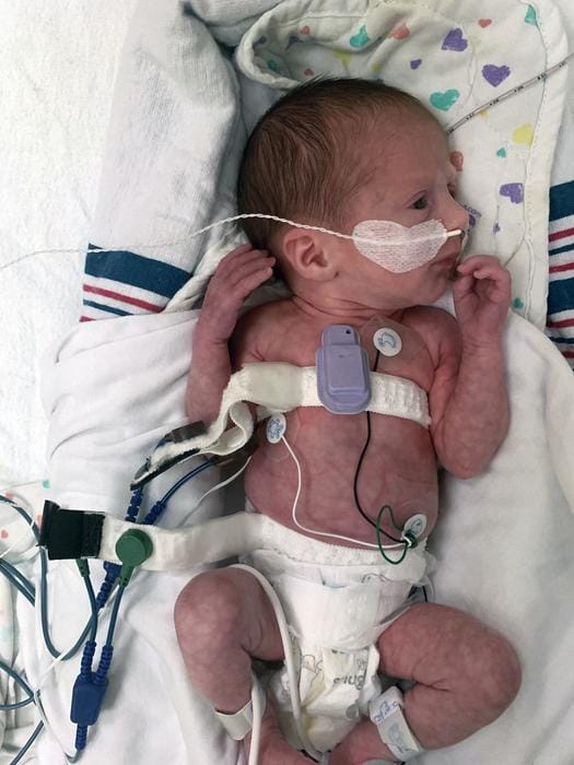
In collaborative studies conducted at the Montreal Children’s Hospital in Canada, health care workers placed the acoustic devices on babies just below the suprasternal notch at the base of the throat. Devices successfully detected the presence of airflow and chest movements and could estimate the degree of airflow obstruction with high reliability, therefore allowing the identification and classification of all apnea subtypes.
“When placed on the suprasternal notch, the enhanced ability to detect and classify apneas could lead to more targeted and personalized care, improved outcomes, and reduced length of hospitalization and costs,” Shalish says in a release. “When placed on the right and left chest of critically ill babies, the real-time feedback transmitted whenever the air entry is diminished on one side relative to the other could promptly alert clinicians of a possible pathology necessitating immediate intervention.”
Mapping a Single Breath
Accompanying the NICU study, researchers tested the devices on adult patients, which included 35 adults with chronic lung diseases and 20 healthy controls. In all subjects, the devices captured the distribution of lung sounds and body motions at various locations simultaneously, enabling researchers to analyze a single breath across a range of regions throughout the lungs.
“As physicians, we often don’t understand how a specific region of the lungs is functioning,” Bharat says in a release. “With these wireless sensors, we can capture different regions of the lungs and assess their specific performance and each region’s performance relative to one another.”
In 2020, cardiovascular and respiratory diseases claimed nearly 800,000 lives in the US, making them the first and third leading causes of death in adults, according to the Centers for Disease Control and Prevention.
With the goal of helping guide clinical decisions and improve outcomes, the researchers hope their new devices can slash these numbers to save lives.
“Lungs can make all sorts of sounds, including crackling, wheezing, rippling, and howling,” Bharat says in a release. “It’s a fascinating microenvironment. By continuously monitoring these sounds in real time, we can determine if lung health is getting better or worse and evaluate how well a patient is responding to a particular medication or treatment. Then we can personalize treatments to individual patients.”
Photo caption: A health care worker places the wearable devices across a patient’s chest to capture sounds throughout the lungs that are associated with breathing.
Photo credit: Northwestern University