
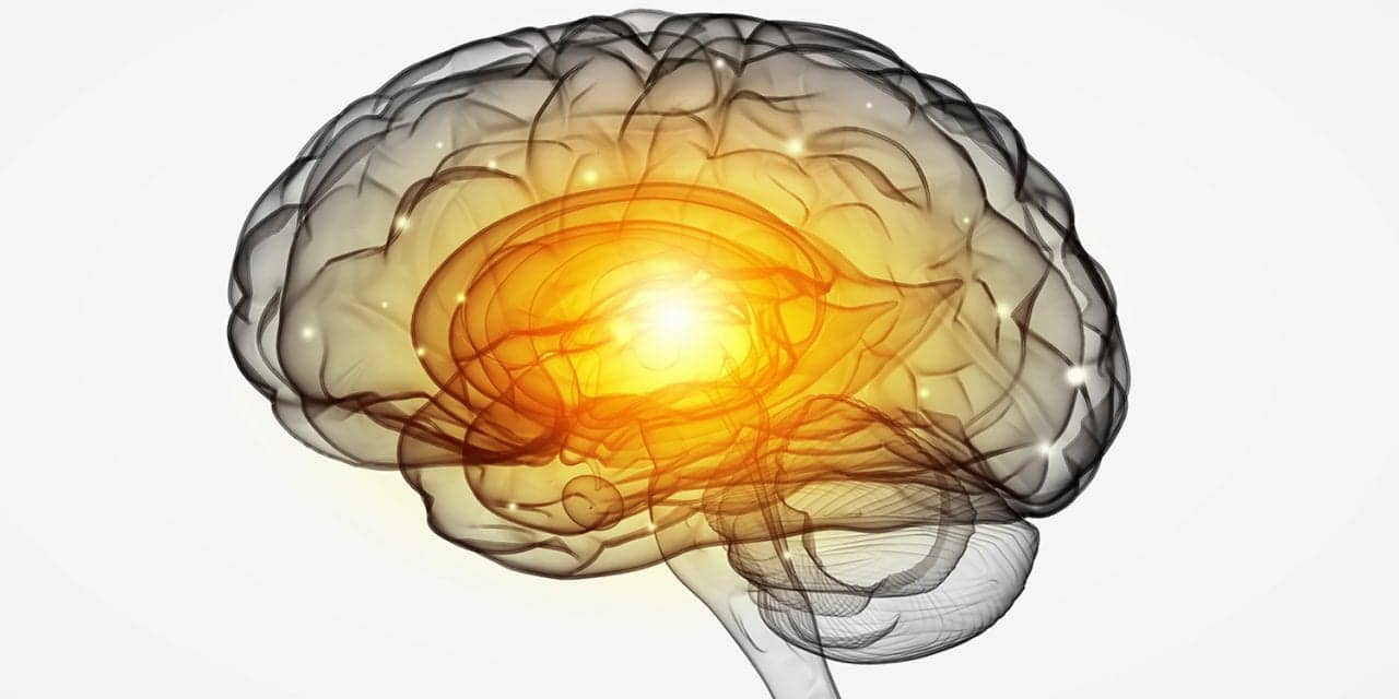
A newly identified group of brain cells actively promotes sleep and coordinates brain rhythms, challenging long-held theories on sleep regulation.
RT’s Three Key Takeaways:
- Active Cortical Role: Researchers found that a rare population of cortical neurons actively drives sleep and synchronizes brain activity, challenging the century-old view that the cortex serves merely as a passive follower during sleep onset.
- Long-Range Signaling: Accounting for roughly 0.2% of cortical neurons, these Sst-Chodl cells project signals across wide areas of the brain to induce slow rhythms characteristic of non-rapid eye movement sleep.
- Clinical Potential: Because this neuronal system has been conserved throughout vertebrate evolution, investigating its function may provide new avenues to understand and manage sleep disorders associated with neurological conditions.
A research team has identified an exceptionally rare population of neurons in the cerebral cortex that actively coordinates brain activity and promotes sleep, according to a study published in Nature.
The discovery challenges a traditional model that has guided neuroscience for nearly 100 years. In that conventional framework, sleep-wake cycles are dictated exclusively by structures located deep within the brain, leaving the cortex—the region responsible for thought, perception, and memory—to passively receive sleep signals. The new findings indicate that the cortex directly participates in initiating and regulating sleep states.
“The traditional view of the cortex is as a passive follower during sleep,” said Renata Batista-Brito, PhD, associate professor of neuroscience at the Icahn School of Medicine at Mount Sinai. “What we found is that the cortex itself contains circuits that can actively drive and synchronize the activity associated with sleep and, when activated, can actually promote sleep.”
Unusual Long-Distance Connections
According to the study, the identified cells, termed Sst-Chodl neurons, make up approximately 0.2% of all neurons in the cortex. While most inhibitory interneurons communicate only with nearby cells, Sst-Chodl neurons extend long-range projections across distant cortical regions, giving the small cell population the capacity to modulate activity across widespread areas of the brain.
In animal models, investigators observed that Sst-Chodl neurons remained largely inactive while mice were awake and alert. However, as the animals entered non-rapid eye movement (NREM) sleep—the phase of rest marked by slowed breathing, decreased muscle tone, and coordinated slow brain waves—these neurons became active as cortical rhythms shifted into slow synchronization.
When the researchers artificially activated the Sst-Chodl neurons, electrical signals across the neocortex slowed and became synchronized, mimicking natural sleep patterns. This targeted stimulation caused mice to fall asleep faster and remain asleep longer, demonstrating that the neurons can actively trigger sleep transitions rather than merely responding to them.
“These neurons don’t simply become active when the animal sleeps,” said Batista-Brito, senior author of the study. “They can actually help drive the transition toward sleep.”
Evolutionary Role and Future Research in Sleep Disorders
Despite their scarcity, Sst-Chodl neurons have been preserved across hundreds of millions of years of evolution, existing in amphibians, reptiles, and mammals, including humans, according to the study authors. This long-term conservation suggests that the cells fulfill a fundamental physiological requirement across species.
“Evolution has held onto these cells for an incredibly long time, even though there are very few of them,” said Batista-Brito. “That suggests they are doing something important. Until now, we knew surprisingly little about what that was.”
The research team also proposed that these specialized neurons may help translate sleep pressure—the biological drive to sleep that accumulates during wakefulness—into actual sleep by directing the cortex to quiet down when fatigue builds.
Disrupted sleep architecture is common in healthcare settings and is frequently observed across neurodevelopmental, neurological, and psychiatric conditions, including Alzheimer disease and autism spectrum disorder. The study authors noted that identifying this cortical circuit creates a new target to determine whether dysfunctional Sst-Chodl neurons contribute to pathological sleep disturbances, which could inform future approaches to sleep medicine and clinical respiratory care.
“Are these neurons functioning normally when sleep becomes disrupted? Could changes in this system contribute to sleep problems in disease?” asked Batista-Brito. “We don’t know yet, but now we have a specific circuit to investigate.”